Category Archives: Clinical Trials

The Case for Low Dose FOLFIRINOX

The 2011 approval of FOLFIRINOX for pancreatic cancer1 was a breakthrough for pancreatic cancer. The one-year survival rates doubled compared to the current best available treatment, gemcitabine. However, concern for toxicity and adverse side effects quickly restricted patients to only the healthiest. In this post, we examine peer-reviewed, published evidence for low dose FOLFIRINOX maintaining effectiveness and reducing patient side effects.

How Treatment Doses are Set

I’ve written previously here how researchers set doses with phase 1 clinical trials. Briefly, in a 3×3 trial design, groups of 3 patients receive a specified dose level and are then assessed for toxic effects. If nothing “bad” happens, the next group of 3 patients receive a slightly increased dose. This cycle repeats until patients experience too many toxic effects. An additional group of patients receives a lower dose to assure the limited toxicity.

The adverse reactions of a handful of patients determines the dosages for all other patients. Note that the treatment effectiveness is not a consideration in setting was is aptly called the Maximum Tolerated Dose (MTD). To be sure, later clinical trials may see additional toxicities and modify the doses or schedule a little, but usually not much. The key idea driving this is that more chemotherapy is better.

Minimum Effective Dose

Ideally, we’d like to discover the Minimum Effective Dose (MED) for any treatment. That could entail large clinical trials with several cohorts taking different doses. To detect small differences in treatment outcomes, we must enroll large groups of patients. Tying up the few patients willing to participate in clinical trials in MED studies would delay development of new treatments.

Is More Always Better?

Smaller doses may be just as effective in treating tumors, and almost certainly result in fewer adverse side effects. Fewer side effects allow patients to stay with treatments longer. There have been a few studies with low dose FOLFIRINOX, so let’s review what they found.

The Evidence for Less FOLFIRINOX

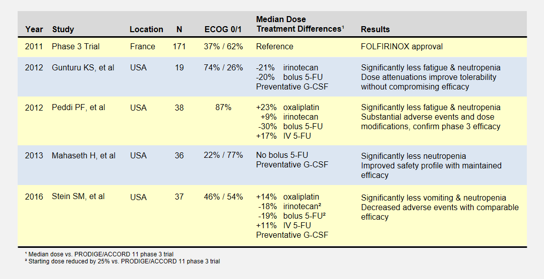
I summarize four studies that report on efficacy and side effects of low dose FOLFIRINOX in metastatic pancreatic cancer. The following table summarizes these studies and the phase III PRODIGE 4/ACCORD 11 approval trial.

Overview of Low-Dose FOLFIRINOX studies.

Overview of Low-Dose FOLFIRINOX Studies

I’ve reported the median dose levels, when available, as compared to the phase 3 FOLFIRINOX clinical trial. Many patients did not start at a full dose, and most had dosage reductions sometime during each study.

About the Studies

The Gunturu KS, et al2 retrospective review included Yale University previously-treated patients from June 2010 to July 2011, documenting their doses, toxicities, and survival results. Reduced doses were not by design, but rather a result of physician discretion. All patients received preventative G-CSF (i.e. neupogen) to prevent neutropenia. This study had a high percentage of healthiest patients.

The Peddi PF, et al3 retrospectively reviewed the FOLFIRINOX experiences of Washington University, the Mayo Clinic, and the University of Wisconsin to compare US “real-world” experiences to the phase 3 clinical trial held in France. Physicians reduced doses at their discretion, not by study design.

Mahaseth H, et al4 retrospectively reported on Emory University’s modified FOLFIRINOX regimen that omitted bolus 5-FU and administered G-CSF to all patients.

Yale University later conducted the Stein SM5, et al prospective study after the promising results in the Gunturu KS study. Starting Irinotecan and bolus 5-FU doses were reduced by 25%, and further at physician discretion. This phase 2 clinical trial (NCT01523457) may provide the most rigorous results.

Patient Responses

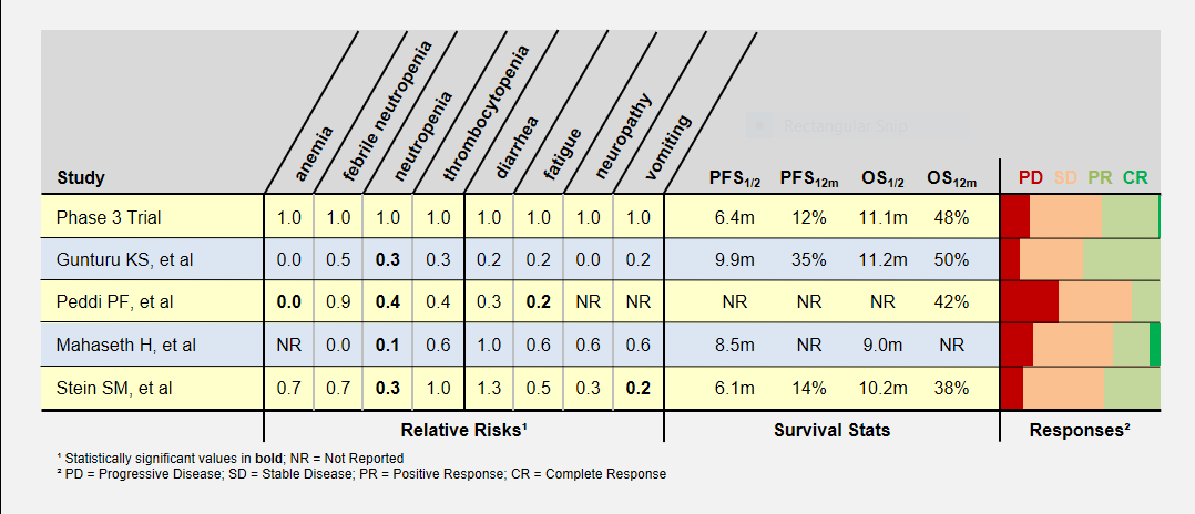
I’ve summarized these study’s adverse patient reactions and tumor response statistics in the table below. Relative Risks of greater and less than 1.0 indicate that the study recorded more or less of a particular event, respectively, compared to the phase 3 clinical trial. As an example, a relative risk of 0.3 means that particular event happened 30% as often as in the phase 3 clinical trial. Bold numbers indicate statistically significant findings (p-value < 0.05). Because of the small study sizes, many promising findings did not reach statistical significance.

Patient responses in Low-Dose FOLFIRINOX studies.

Patient Responses in Low-Dose FOLFIRINOX Studies

Progression-Free (PFS) and overall survival (OS) were similar in all studies (the accurate statement is that they were “not found to be different”). Toxicity levels were almost uniformly lower in all low dose FOLFIRINOX studies.

Colored bars represent tumor responses to treatment, with width corresponding to patient counts. Depending upon an individual’s treatment goals, you may want to know different results. For instance, a patient needing to shrink a tumor for surgery may want to look at the PR+CR result. A patient desiring long-term stability may want to minimize the PD result.

Blood-Related Adverse Events

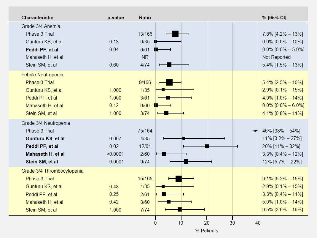
The table below contains more detail and 95% confidence intervals on blood-related adverse events. Studies with fewer participants have less certain results and wider confidence intervals. For adverse event analysis, I included both locally advanced and metastatic patients from each study.

Blood related adverse events of Low-Dose FOLFIRINOX Studies.

Blood Related Adverse Events in Low-Dose FOLFIRINOX Studies

Most adverse events for lower dose FOLFIRINOX were less than the baseline phase 3 clinical trial. However, except for neutropenia, sample sizes or adverse effects were too small to show statistical significance.

Note that the phase 3 clinical trial’s neutropenia rates were especially high. Three studies (Gunturu, Mahaseth, and Stein) used C-GSF for all participants which likely reduced the rate of neutropenia. The Peddi study used C-GSF at higher rates than the phase 3 trial, also reducing the neutropenia rates.

Non-Blood Related Adverse Events

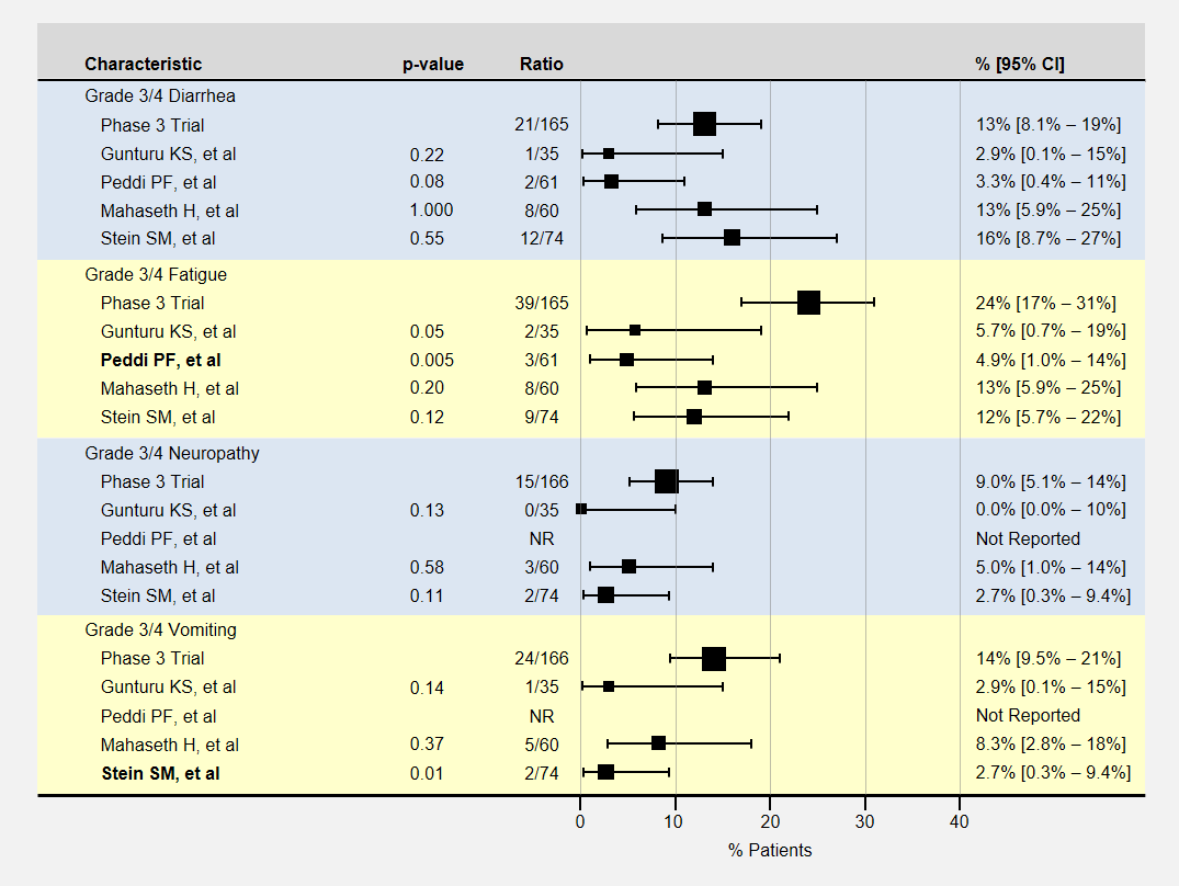
The table below contains more detail and 95% confidence intervals on non-blood-related adverse events. For adverse event analysis, I included both locally advanced and metastatic patients from each study.

Non-blood related adverse events of Low-Dose FOLFIRINOX studies

Non-Blood Related Adverse Events in Low-Dose FOLFIRINOX Studies

Here again, most adverse events for lower dose FOLFIRINOX were less than the baseline phase 3 clinical trial, with sample sizes usually too small to show a statistically significant effect. The Peddi study had significantly less fatigue, and the Stein SM study with a 25% reduction in the initial dose of irinotecan and bolus 5-FU had significantly less vomiting.

How Low Can We Go?

Let’s take a look back at the phase 1 dose escalation trial for FOLFIRINOX6. The study establisged a MTD for oxaliplatin and irinotecan at 85mg/m2 delivered over 120 minutes and 220mg/m2 delivered over 90 minutes respectively. The reduced the recommended level to 85/180 mg/m2 when they realized the cumulative effect would not allow patients to maintain a 2 week schedule.

The dose escalation started with lower oxaliplatin/irinotecan doses as shown in the following table. Important to this discussion, almost every dose level showed anti-tumor activity – including complete responses at the two lowest levels. Participants experienced no dose-limiting toxicities at the three lowest dose levels.

Patient responses in phase 1 FOLFIRINOX dose escalation study

Patient Responses in Phase 1 FOLFIRINOX Dose Escalation Study

The primary goal to determine the dose-limiting toxicity, not effectiveness, opened enrollment to 41 patients with 8 different types of cancers. Of the 6 pancreatic cancer patients, 1 enjoyed a complete and another a partial response (dose levels not specified).

Summary

These peer-reviewed, published studies proved evidence of low dose FOLFIRINOX efficacy similar to the phase 3 trial results. Positive or negative differences in progression-free or overall survival rates cannot be seen with the small studies thus far.

Even with these small study sizes, we have statistically significant evidence of a reduction in toxicity with the low dose FOLFIRINOX regimens.

In the FOLFIRINOX dose escalation trial, patients at the two lowest oxaliplatin/irinotecan levels recorded complete responses with no limiting side effects.

Low dose FOLFIRINOX is better tolerated with significant anti-tumor activity. Oncologists should consider this regimen as an option for patients desiring efficacy but unwilling to endure severe toxic events. Patient experiences with low dose FOLFIRINOX will produce more retrospective studies that will help pinpoint a most effective dose.

I propose that oncologists start patients with low dose FOLFIRINOX and in the absence of severe side effects, increase the dose. The first rounds are often the most difficult as a patient must quickly learn how to deal with chemotherapy-induced nausea and fatigue in addition to their new cancer diagnosis.

References

[1]Conroy T, et al. (2011 May 12) “FOLFIRINOX versus gemcitabine for metastatic pancreatic cancer”. N Engl J Med 364(19):1817-25 PMID: 21561347.

[2]Gunturu KS, et al. (2013 Mar) “FOLFIRINOX for locally advanced and metastatic pancreatic cancer: single institution retrospective review of efficacy and toxicity”. Med Oncol 30(1):361 PMID: 23271209.

[3]Peddi PF, et al. (2012 Sep 10) “Multi-institutional experience with FOLFIRINOX in pancreatic adenocarcinoma”. JOP 13(5):497-501 PMID: 22964956.

[4]Mahaseth H, et al. (2013 Nov) “Modified FOLFIRINOX regimen with improved safety and maintained efficacy in pancreatic adenocarcinoma”. Pancreas 42(8):1311-5 PMID: 24152956.

[5]Stein SM, et al. (2016 Mar 29) “Final analysis of a phase II study of modified FOLFIRINOX in locally advanced and metastatic pancreatic cancer”. Br J Cancer 114(7):737-43 PMID: 27022826.

[6]Ychou, et al. (2003 Mar) “An open phase I study assessing the feasibility of the triple combination: oxaliplatin plus irinotecan plus leucovorin/ 5-fluorouracil every 2 weeks in patients with advanced solid tumors”. Ann Oncol 14(3):481-9 PMID: 12598357.

Cancer Models

Driving treatment decisions on 25 Sep 2015

Cancer models are used by scientists to define and better understand cancer. They describe the rules cancer follows – how it starts, grows, metastasizes, and ultimately how it can be killed. Reviews of their cancer models generate new treatment proposals that the models indicate should be successful.

Cancer models are driving the direction of cancer funding, research, prevention and treatment. Faulty cancer models lead to research producing ineffective treatments. Some researchers say that is happening now.

Cancer Models Example

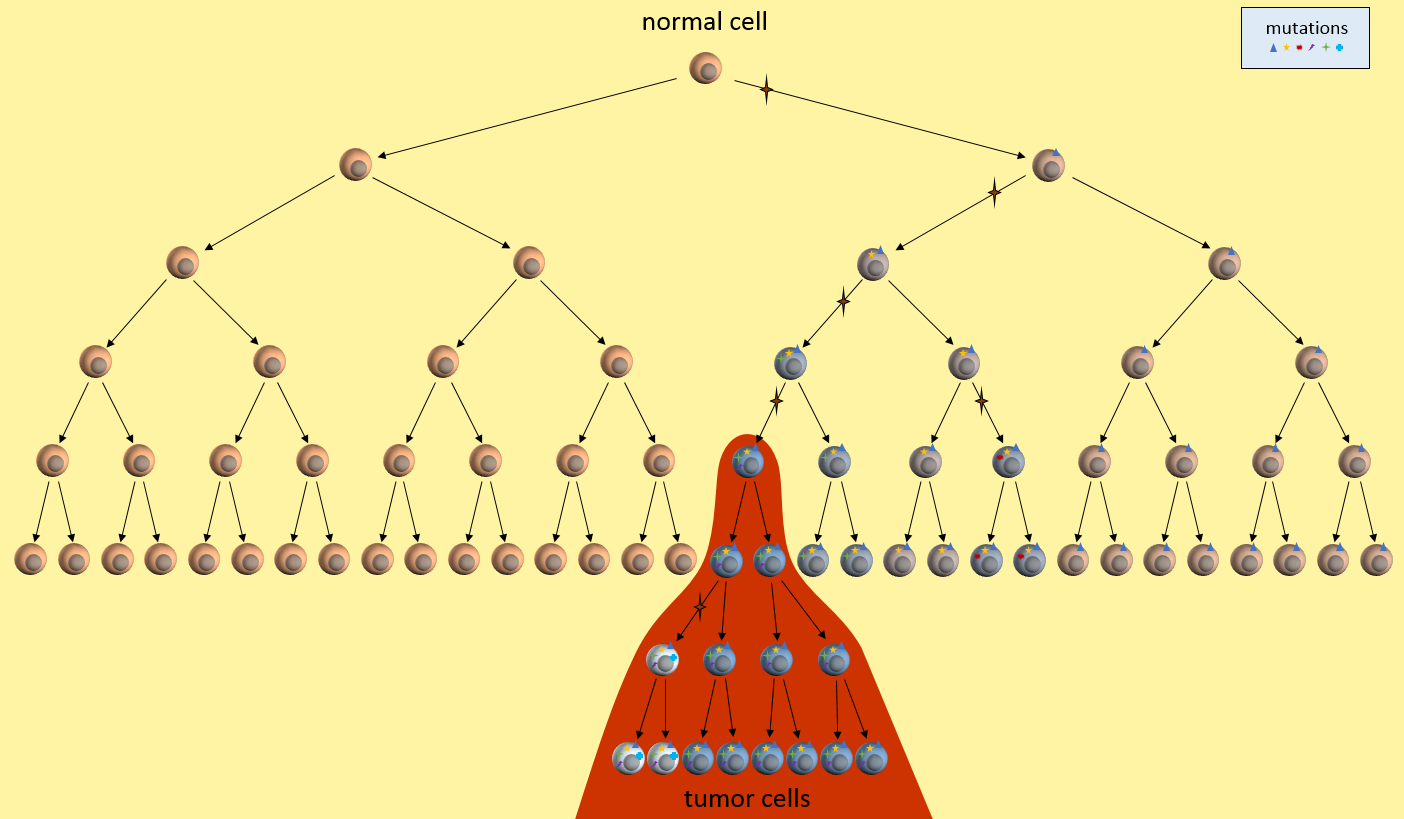
Let’s look at one example of a simple cancer model. It’s just a picture that describes how a normal cell might progress to a cancer cell and then a tumor. In this model, each time the cell divides, a mistake in the replication process results in a mutation (indicated by the star symbol) in one of the two daughter cells. After enough divisions with accumulated mutations, a cancer tumor cell results (highest cell in the red area). All subsequent daughter cells are cancer cells and additional mutations keep happening. The tumor ends up containing cells with varying kinds of mutations. However, the mutations present in the first tumor cell remain present in all daughter cells. These are called driver mutations1, because they drove the initial cancer and will be present throughout the entire tumor.

Model of Mutation Accumulation in the Clonal Evolution Model

Let’s just take this simple model, and ask ourselves some questions. What are this model’s weaknesses? What treatment decisions might this model recommend?

Testing the Model

First, let’s look at what our model implies about cancer growth. Does it fit with observations from real-world patients?

Mutations Only During Replication?

What does our model tell us about cancer growth? For instance, is it reasonable that mutations only happen at during replication? Well, we seem to know that cells need about 20-30 mutations before they can become cancerous. We also know that normal cells can only replicate about 40 to 60 times before their telomere length is too short to allow any more replications. Given this, and DNA’s ability to repair itself, we might have to adjust our model to allow more than one mutation per replication, or allow mutations to happen without replication, or grow longer telomeres. We might design experiments to determine which of these is more correct and then update the model to reflect the new findings.

Are There Nearby Pre-Cancerous Cells?

Our model also seems to indicate that there will be many cells nearby with some mutations, but not enough to yet be cancerous, or pre-cancerous. Is this something that a pathologist can determine? Can we separate out nearby normal-looking cells and genetically sequence them to see if they have some mutations but not all?

Timeline Of Cancer Development?

We might observe that our model seems to require quite a few generations of replications before the first cancerous cell develops. Where to all these cells go? What does this tell us about how quickly cancer could develop? What ages cancers might develop? Does this fit in with the childhood leukemia? Does it fit in with pancreatic cancer? Do we need different models for these cancers? Which cancers does it fit?

Cancer models raise many questions like these that drive directions of research that demand funding. Answering these questions allow us to tweak the model to be more accurate. The more accurate the model, the better its predictive power for new treatments.

Cancer Model Development Flowchart

Cancer Model Development Flowchart

Refining the Model

After we’ve tested the model and experimentally determined new facts, we can update the cancer model. The updated model would be tested again, continually refining the model.

Using the Model

Once we’re happy that the cancer model is mostly reflecting reality and is able to answer basic questions about cancer’s behavior, we are ready to put this model into practical use.

Treatment Decisions

What does our model tell us about treatment decisions? For one, this model seems to indicate that there are certain mutations that will be present in all daughter tumor cells. Examine those nearby pre-cancerous cells. Do they hold the key to uncovering these cancer driver mutations1? If we can find these driver mutations and target them with treatments, we might be able to kill off the entire tumor. This is the idea behind the NIH’s Precision Medicine Initiative.

Tumor Heterogeneity

Our model also says that many mutations will only be present in a part of the tumor – not the entire tumor. See those white and blue cancer cells in the model? They represent tumor cells with different sets of mutations. If we treat only for the white cells’ mutations, the blue cells might continue to grow. This observation fits current patient outcomes.

Early Detection

What does our model say about early detection? Our model indicates that precancerous cells will be around for a while before the cancerous cells develop. It there some way we might be able to detect these pre-cancerous cells? Perhaps they affect the environment around them or put out markers that might be picked up in the bloodstream? This model might drive the development of another model that looks at these issues in the cellular environment.

(Re)-Refining the Model

The cancer model is always being updated to reflect the best knowledge of how cancer works. This simple model really just deals with cancer at a very high level, but even so, it reveals directions for research, early detection, and treatments. Continual refinement of even this simple model brings forth new ideas to be tested, further feeding the models.

Summary

Don’t think that there is some “master” cancer model somewhere that all researchers work from. Each group has its own models developed from their own experience, lab tests, ideas from other groups, etc. These cancer models describe some aspect of cancer as it is understood by the local research team.

In the next posts, I’ll describe two current cancer models. The clonal evolution model2 that has been driving research and funding decisions for several decades. And the cancer stem cell model3 that is not widely accepted but is gaining traction. Which cancer model is (more) correct has a profound effect on patient treatment choices.

Our lack of progress could mean we need to take another look at our basic assumptions, the cancer model, and develop a new model that describes not only how cancer starts, grows, and metastasizes, but also accounts for why we’ve been failing in the “war on cancer”. Each model will undergo continuous revision and tweaking based on new tests to improve their accuracy.

References

[1] Stratton MR (9 April 2009). “The cancer genome”. Nature 485(7239):719-24. PMID: 19360079.

[2] Nowell PC (October 1976). “The clonal evolution of tumor cell populations”. Nature 194(4260):23-8. PMID: 959840.

[3] Soltysova A, Altanerova V, et al. (2005). “Cancer stem cells”. Neoplasma 52(6):435-40. PMID: 16284686.

What Researchers Know and You Don’t

An explanation for what’s driving drug development

In one of the most highly cited cancer papers, The Hallmarks of Cancer1, researchers Doug Hanahan and Bob Weinberg described six common traits of cancer. Their 2011 update, Hallmarks of Cancer: The Next Generation2, added two more traits for a total of eight “hallmarks” of cancer. They also describe an additional two “enabling characteristics” that are not necessary, but if present, hasten the cancer process.

In these papers, they hypothesize that eight specific functions of normal cells must be impaired for them to become cancer cells. A cell could develop these impairments over time, in any order, but only after developing all or most of them could it become a cancer cell.

The hallmarks listed in these papers are understood by every cancer researcher and referenced by thousands of research papers. This is what researchers know and you don’t – yet.

What Does This Mean For Patients?

  • The time needed to accumulate all these impairments explains why cancers are more likely as we age.
  • The requirement to have all these impairments together explains why cancers develop so rarely.
  • Inheriting a mutation of an enabling characteristic explains why hereditary cancers develop in younger people. In essence the deck is stacked against them.
  • A treatment targeting any one of these hallmarks would prevent a tumor from developing.

Implications for Cancer Treatment

The last item above explains a lot about the current direction of cancer drug development. If you were to repair any of these functions, you could potentially stop cancer. Targeted treatments being developed today attempt to repair one or more of these traits in an effort to halt cancer growth.

For example, one hallmark is that cancer cells evade detection by the immune system. This means that cancer cells have developed some way to keep the immune system from recognizing the cancer cells as undesirable. Current immunotherapy drugs are trying to “fix” the immune response to recognize the cancer cells. In pancreatic cancer, treatments currently in later-stage clinical trials that target this hallmark include GVAX vaccine (Johns Hopkins & Aduro BioTech), Algenpantucel-L (NewLink Genetics), CAR T-cell therapy (U of Penn)4.

Patients should note that when cancer cells are targeted in a specific hallmark, they will often develop an alternate method of re-impairing the hallmark function and continue growing as a tumor. For example, there are many ways of deactivating the immune system. Fixing one part of the immune response usually leads to the cancer cells developing an alternate method to halt the immune response. Cancer cells’ ability to continue mutating as they divide means they can eventually stumble on an alternate mechanism. I imagine the tumor as a massively parallel computer that can try thousands of mutation experiments simultaneously where only needs one to succeed at foiling the treatment. Researchers can only target a specific one of these mechanisms at a time.

In the future, perhaps we will combine these therapies in a multi-pronged attack on cancer cells such as is proposed for NSCLC5? Or alternating between two effective treatments to keep cancer from developing resistances? I suspect that this process is at the beginning of a long road.

Hallmarks of Cancer

Here are the hallmarks of cancer in plainer English. The explanation in FutureLearn‘s MOOC Cancer and the Genomic Revolution6 was well presented.

Hallmark1, 2Example Therapeutic Targets2
Growth signals stuck ONEGFR Inhibitors
Ignore anti-growth signalsCyclin-dependent Kinase Inhibitors
No "kill" switchProapoptotic BH3 Mimetics
Unlimited replicationTelomerase Inhibitors
Compels new blood suppliesVEGF signaling Inhibitors
Migrate to other organsHGF/c-Met Inhibitors
Energy production using little O2Aerobic Glycolysis Inhibitors
Deactivate immune systemImmune Activating anti-CTLA4 mAb
Enabling Characteristic2
Easier MutationPARP Inhibitors
Favorable inflammation environmentAnti-inflammatory drugs

I’ll try describing the hallmarks of cancer with an automobile factory analogy. In this analogy, cancer cells are automobiles and uncontrolled growth is accomplished by means of the factory building these autos. These automobiles are often defective, unsafe, but they’re practically free and plentiful. But because of the random defects in these autos, many of them don’t even work at all. But some do and they can have scary defects.

In a more correct analogy, each tumor cell is its own factory producing more defective cars, but we’ll stick with the one factory for now.

The heading for each Hallmark is followed by the name from the original paper in italics and in parentheses. I think you’ll understand why I’ve interpreted them.

Always ON Growth Signals (Sustaining Proliferative Signaling)

One attribute of cancer cells is that they are always replicating, growing the tumor larger and larger.

In our automobile factory, the assembly line never stops. It is working three shifts every day of the year, putting our defective cars out on the road.

Ignore Anti-Growth Signals (Evading Growth Suppressors)

Cancer cells ignore signals that neighboring cells send saying that there’s enough of them and they should stop dividing.

Our dealerships can’t even store all the cars we’re making. They are saying, “Stop! We don’t want any more of your cars!” But their pleas fall on deaf ears. This factory keeps producing cars.

No Kill Switch (Resisting Cell Death)

Cancer cells are made with lots of mistakes in their DNA. Normally, if those defects cannot be fixed, the cell is instructed to kill itself.

Our workers are pretty sloppy and we’re always producing cars with random mistakes. But in our factory, we have no quality control so that all cars are shipped. A lot of them don’t work at all. But some of them do and have some pretty scary attributes. In some, the brakes don’t work. In others, the accelerators are stuck on. In still others, airbags won’t deploy. All defects that should be caught and cause these autos to never see the light of day. But we’ve got no quality control so every car ships.

Unlimited Replication (Enabling Replicative Immortality)

Cancer cells can replicate themselves almost without limit. Normal cells can only replicate up to about 20 times. Their telomeres are shortened by every replication, but cancer cells have found a way to replicate without shortening their telomeres.

In our factory, the robots, machines and tools never wear out or break down. We can produce cars almost without limit.

Compel New Blood Supplies (Inducing Angiogenesis)

Cancer tumors grow very rapidly but still need to be supplied with blood and nutrients. These cells send out signals tricking arteries into branching into the growing tumor.

Our factory managers have bribed their suppliers to provide all the raw materials we need to continue making our cars. They’re not paying them properly and we’ve resorted to trickery to obtain our supplies.

Migrate to Other Organs (Activating Invasion & Metastasis)

Eventually cancer cells develop a way to metastasize. As an example, a cluster of perhaps 100 cells detaches from the tumor, entering the blood stream and gets caught by the liver as it tries to filter out impurities. There it lodges and starts to grow inside the new organ.

This automobile factory becomes so successful at producing cars that the managers decide to open a new factory in another state or country.

Energy Production Using Little O2 (Deregulating Cellular Energetics)

Cancer cells use sugar in a different way than normal cells. For not well-understood reasons, cancer cells have developed a less efficient process to make energy that also uses less oxygen.

Our factory needs lots and lots of energy to make these cars, 24×7. We have access to the same power source that any factory would have, but we also have solar and wind power to supply our energy demands. We’ve developed alternate sources of energy.

Deactivate the Immune System (Avoiding Immune Destruction)

Cancer cells are really normal cells that have mutated to have all the above attributes. The immune system does not recognize them as foreign invaders because they really aren’t – they came from inside our own bodies. Even if the immune system did start to attack, cancer cells have figured out how to distract the immune cells.

Our factory has figured out how to avoid government regulations. We keep producing defective cars, cheating our suppliers, have unapproved power sources, and clearly run afoul of labor laws, but no one comes in to shut us down. Perhaps the factor owners have paid “protection money” to keep regulators looking the other way?

Summary

Articles like The Hallmarks of Cancer provide an insight into the thinking of researchers. Understanding the Hallmarks can help direct you to promising treatments and understand why we’re not at a curative treatment stage yet. Highly mutative tumor cells can bypass any single drug we develop. Eventually we may develop enough targeted treatments that used together will halt cancer growth.

To me, it reinforces the idea that surgical removal is still the best and only pancreatic curative option. Working towards that option will be my primary goal.


References

[1] Hanahan D, Weinberg RA (January 2000). “The Hallmarks of Cancer”. Cell 100 (1): 57–70. doi:10.1016/S0092-8674(00)81683-9. PMID 10647931

[2] Hanahan, D.; Weinberg, R. A. (2011). “Hallmarks of Cancer: The Next Generation”. Cell 144 (5): 646–674. doi:10.1016/j.cell.2011.02.013. PMID 21376230

[3] Scowcroft, Henry (2010). Science blog: http://scienceblog.cancerresearchuk.org/2010/11/10/ncri-conference-the-hallmarks-of-cancer/

[4] Cancer Research Institute’s Pancreatic Cancer web page (accessed 7 Aug 2015) http://www.cancerresearch.org/cancer-immunotherapy/impacting-all-cancers/pancreatic-cancer

[5] Turke, Alexa B, et al. (January 2010). “Preexistence and Clonal Selection of MET Amplification in EGFR Mutant NSCLC”. Cancer Cell 17 (1): 77-88. doi:10.1016/j.ccr.2009.11.022. PMID 20129249

[6] FutureLearn MOOC, Cancer and the Genomic Revolution, University of Glasgow. https://www.futurelearn.com/courses/cancer-and-the-genomic-revolution/

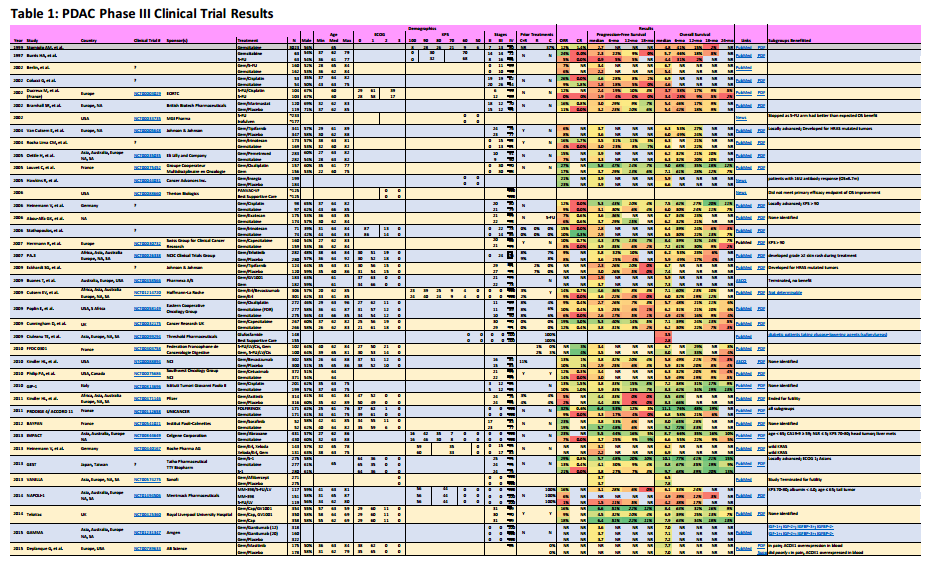
Phase 3 Metastatic Pancreatic Adenocarcinoma Clinical Trial Results

Overview

Metastatic pancreatic adenocarcinoma patients (PDAC) don’t have a lot of good options. In this post, I’ve summarize the completed chemotherapy-based phase III trials for metastatic patients, providing links to publications, outlining the survival statistics, and identifying key subgroups that may have benefited from the treatment.

From a patient’s perspective, not much progress has been made on new treatments. It’s not for a lack of trying. Over 90% of phase 3 clinical trials result in no added benefit.

However, I believe that these treatments are effective on a minority of patients. The data used to measure effectiveness (like median OS) ignores outstanding responses to less than half the patients. I propose that looking at longer term survival rates gives clues to treatments effective on a minority of patients. Further digging into the results by researchers can uncover reasons behind the effectiveness and help us match treatments to patients.

Key Points for Patients

  • Several treatments available. We need to better match the right treatment to the right patients. Some studies have published information about subgroups that benefited substantially from treatment in a trial. This can help you choose whether that treatment is right – or wrong – for you.
  • A patient’s treatment goals should determine which trial results are important.
  • Directly comparing results between trials is misleading due to the fact that patient selection is key to the results and each trial has its own selection criteria.
  • Results in italics are values I’ve read from published graphs, not specifically published by the study paper.
  • Results in bold are for treatments often used for pancreatic cancer.

Observations

In reviewing the information to put together this information, I’ve formed some interesting opinions that I’ll share.

First, I should state that the results provided here are for the actual patients that were under clinical trial. Your results will almost certainly not be “typical”. There are uncertainties in the outcomes that are hopefully minimized because these are phase 3 clinical trials with large numbers of patients. One of my goals here is to identify the best performing treatments that can maximize your outcomes. If we can identify some treatments that work best for patients in your particular situation, this posting has done its job.

This summary lists very little about side effects of the treatments. For some, this can also be a big factor in a treatment decision. If you are in poorer health, try looking for the best performing trials that included patients with poor health (ECOG 2/3 or KPS≤70).

In the data listed, numbers provided directly from the clinical trial results are listed in normal fonts. Rates in Italics have been interpolated from graphs provided by the clinical trials publications.

Click on the following picture to show a PDF file with the results.

Phase 3 PDAC Trials (small)

Pancreatic Adenocarcinoma Phase III Clinical Trials

Subgroups Benefited

I think this is the most useful information available from the data I’ve collected.

Some clinical trial reports are superior to others. Take for instance, the reports for FOLFIRINOX and Gemcitabine/Abraxane. Both of these reports include what are called forest plots to identify subgroups of patients that did well on one treatment versus the other.

The FOLFIRINOX forest plot is here (I provide the link because I’m unsure whether I can legally embed the figure, which might be clearer). For each subgroup listed, a hazard ratio has been computed (square box), with 95% confidence intervals shown (horizontal line). Subgroups listed that have the entire confidence interval (horizontal line) to the left of 1.0 (which is just about everything) performed better under FOLFIRINOX than Gemcitabine. This kind of information can be vital to patients who belong to one of these subgroups when they’re trying to decide on a particular treatment.

In the column Subgroups Benefited, I try to list groups indicated within these reports that did benefit from one treatment arm. Sometimes, a follow-on report has the information and I have provided hyperlink in the column for these cases.

ORR

This is the percentage of patients that had at least a 20% reduction in the size of their tumor. FOLFIRINOX has the best ORR at 32%. Understand that 2/3d’s of treated patients did not have their tumors shrink using the best treatment available.

The key is finding out why these patients had their tumors shrink. Are you more likely to belong in this group? Again, this is why the subgroup analysis is important.

CR

If your treatment goal is to remove all metastases to enable a chance at surgery, CR (Complete Response) is your result of interest. These are pitifully small numbers. But what this tells me is that some small subgroup of patients do respond remarkably well to some treatments. One example might be BRCA2 mutation patients that respond extraordinarily well to platinum-based treatments [reference needed].

PFS

If your treatment goal something like living as long as possible without worsening symptoms., this might be your series of results to look at. Often, only the median is reported, but I favor looking at the 12- or 18-month PFS percentages. This tells me how many patients had a sustained response to the treatment. Their tumor shrunk and they’re tolerating the treatment.

OS

This is usually a clinical trial’s primary end statistic. Median numbers are what researchers compare but I find them largely irrelevant to patients. I want to see the 18- and 24-month OS percentages. These are the long-term survivors. Compare the PFS and OS percentages and know what fraction of survivors are still benefiting from the treatment.

Clinical Trial Progress

Often we patients are frustrated by the lack of progress towards more effective treatments. Here I’ve documented over 38 different chemotherapy phase III clinical trials since 2002 (I’m certain there are more). Only four of these trials led to approval, a success rate of about 10%.

In general, these are drugs that had successful phase II results and then failed during phase III. First, it helps to demonstrate that there is still significant effort being put into new treatments. But second, how many very promising treatments fail to show patient benefit.

Interesting Results

When a trial fails to show benefit, often that is the end of the road. However, sometimes the researchers see interesting things buried in their data. Here are a few I’ve uncovered.

Gemcitabine/S-1

Here’s a stellar performer that I think should be approved pronto! For Asians. And therein lies one of our problems with the clinical trial system. It is stacked in favor of Caucasians. This combination works well and is well tolerated by Asians only. Caucasians – not so much.

Gemcitabine/Cisplatin & Gemcitabine/Oxaliplatin

The trial results for these platinum-based chemotherapies did not identify any subgroups that performed well, but it has since been recognized that many patients with a wild-type BRCA2 mutation (about 5-10% of all pancreatic cancers) have responded quite well (2009 Dramatic Response; 2011 Complete Response; 2012 Complete Response; 2014 Meta-analysis). I credit my complete response to Gemcitabine/Cisplatin because of my BRCA2 mutation.

Gemcitabine/Masitinib

According to the traditional measurement criteria (median PFS and/or OS), this trial failed miserably. A closer reading of the results (kudos researchers!) identifies that PDAC patients with overexpression of ACOX1 in their blood did much better than others (Figure). What’s less clear is whether this finding also says that patients with an overexpression of ACOX1 in their blood should avoid Gemcitabine monotherapy.

Gemcitabine/Ganitumab

Another research report (behind a paywall, so not fully reviewed by me) of a failed clinical trial. But a later closer analysis of the results in another paper delivers news of biomarkers (IGF-1+; IGF-2+; IGFBP-3+; IGFBP-2-) that identified a subgroup of patients that survived 3X longer than the others.

Gemcitabine/Erlotinib

Erlotinib (aka Tarceva) was expected to target tumors that overexpress EGFR, like pancreatic cancer. While the EGFR status of patients did not seem to affect the outcomes, development of rashes did. The more severe the rash, the better the outcomes (Figure). In fact, some oncologists are increasing the doses of erlotinib until a rash develops.

Summary

There have been a surprising number of phase III clinical trials for metastatic pancreatic adenocarcinoma. Most do not pan out. And researcher responsibilities should not stop there. It appears unlikely we will soon find a treatment that works for most pancreatic cancer tumors. More effort is being made to identify subgroups that respond well, but that information is not widely disseminated to patients. Hopefully this blog entry will bridge some of that gap.

Immunotherapy of Pancreatic Cancer – a Webinar

WebinarImmunotherapy of Breast and Pancreatic Cancers
PresenterDr. Elizabeth Jaffee, Johns Hopkins
PublisherCancer Research Initiative
Links[Webinar] [Publisher]

Overview

In this blog post, Immunotherapy of Pancreatic Cancer – a Webinar, I’m annotating a webinar for information I think is useful to pancreatic cancer patients. My goal is to make the information more accessible. Towards that end, drugs mentioned are in bold font, and links are provided to additional information, such as research papers, research results, and clinical trials mentioned in the webinar. The webinar information is presented in a table, ordered by time index, with a brief description of the content from the webinar. My interpretation of Dr. Jaffee’s presentation could be incorrect in some areas, so your own healthcare professional’s interpretations should supersede mine.

The Jun 13, 2013 webinar annotated in this posting is from the Cancer Research Institute (CRI) Breakthroughs in Cancer Immunotherapy webinar series. The CRI is a “nonprofit organization dedicated exclusively to harnessing the immune system’s power to conquer all cancers”.

The presenter, Dr. Elizabeth Jaffee, MD [papers], is a Johns Hopkins medical oncologist specializing in vaccine therapies for pancreatic cancer and other solid tumors.

Readers of this blog should note that I am a participant in one of Dr. Jaffee’s immunotherapy clinical trials (NCT01088789) for resected pancreatic cancer patients, receiving a single dose of cyclophosphamide and the GVAX vaccine at each treatment session.

Key Points for Patients

  • Immunotherapy of pancreatic cancer is currently only available through clinical trials
  • Immunotherapy drug targets can be applied to more than one type of cancer
  • This webinar focuses largely on the GVAX vaccine that Dr. Jaffee helped develop
  • Some immunotherapy clinical trial results are presented
TimeDescription
2:02Introduction of Dr. Elizabeth Jaffee
3:31Aduro Biotech has licensed the GVAX and Listeria vaccines
4:58List of technical advances that enabled recent immunotherapy progress
5:44Ipilimumab (Yervoy), FDA approved for melanoma, targets the CTLA-4 protein receptor on the immune T cell so it can recognize the cancer cell as foreign. Believes that it can also work in pancreatic cancer (see Time Index 24:43 and NCT00836407).
7:01PD-1/PD-L1 mAb (monoclonal antibodies), similar to Ipilimumab, inhibits signals on T cells that prevent them from fighting cancer
7:31Listing of new immunotherapy treatments
7:55The targets on immune cells are not tumor type [site of origin] specific. The same principles can be applied to other cancers, such as pancreatic cancer. [DD: The idea of treating cancers by their genetic differences rather than site of origin is being discussed as a major treatment change - see: targeted therapies]
8:31Biomarkers are being developed to identify patients that will respond to a particular immunotherapy. [Paper: Lymphocyte Counts in GVAX]
8:55Explanation (with graphic) of how pancreatic cancer develops. Describes genetic and immune system changes over time [Figure].
9:17Describes a gradual progression of genetic changes. In pancreatic cancer, one of the first changes is a mutation of KRAS.
10:12The tumor cells induce some bad changes in the immune response that help the cancer to grow.
11:26Microscopic view of resected patient's pancreatic cancer showing tumor cells and stroma. The cancer cells attract the regulatory T cells (Tregs, the wrong kind of immune cells) [DD: these Treg cells prevent the right kind of immune cells from getting to the cancer cells. Later GVAX clinical trials added cyclophosphamide to engage the Treg cells and allow the right immune cells to get into the tumor].
12:14Slide and explanation of the tumor's immune-related environment. The CD8+ T cells (a type of effector T cell that has the CD8 protein on its surface) are the ones we need to attack the tumor.
13:14The dendritic cell (DC) can active or suppress T cells in attacking cancer cells. Drugs that inhibit signals such as CTLA-4 and PD-1 keep the dendritic cells from stopping the T cells from doing their work.
14:33Slide showing how the GVAX vaccine and cyclophosphamide work together to bring CD8+ T cells to tumor cells. [2008 Paper][2009 Paper]

  1. The GVAX vaccine containing tumor cells genetically modified to produce GM-CSF is injected just below the skin. The body's dendritic cells detect the GM-CSF emitted by these tumor cells as foreign.

  2. The dendritic cells travel to the lymph nodes where they activate T cells to recognize this new foreign invader.

  3. The newly activated CD8+ T cells leave the lymph nodes in search of tumor cells.

  4. In earlier clinical trials without cyclophosphamide, the Treg cells would intercept the CD8+ T cells before reaching the tumor cells

  5. After recognizing what was happening, a low dose cyclophosphamide was added to the regimen to engage the T reg cells and allow the CD8+ T cells to get to the tumor cells.[Paper]

15:35Mesothelin is one of the proteins on the tumor's surface that is targeted by the immune cells. Mesolthelin levels on the tumor help predict disease-free survival time (DFS) [2004 Paper][2008 Paper][2011 Paper]. [DD: The explanation is not clear here, but I think the finding correlating mesothelin levels on the tumor and DFS is for patients in general (no vaccine)]
17:01Results of 60 patient clinical trial [NCT00084383, Johns Hopkins, NCI] using GVAX (alone) [Paper] that observed improved DFS and that long-term survivors developed an immune response to mesothelin [Figure]. It takes at least 4 vaccine treatments to get to the point of high immune response to mesothelin.
18:27GVAX vaccine clinical trial [NCT00727441, Johns Hopkins, NCI] where first treatment is given 2 weeks prior to surgery. The removed tumor is examined for effects from the vaccination. Lymphocytes activated against the cancer are seen under the microscope [Paper]. [DD: Why aren't more studies done like this? Seeing the effect in a human with cancer is optimal!]
19:44The resected patient's immune cells (lymphoid aggregates) that were attacking the tumor cells are genetically sequenced to see what they were targeting on the cancer tumor cells.
21:04Case study of clinical trial participant that then joined a follow-up/boost study [NCT01088789, Johns Hopkins] whose CT and PET scan appeared to show a recurrence, despite the patient feeling great. Patient went to surgery and the resected tissue showed inflammation with immune cells and no tumor. Theorized that the tumor was coming back and the immune system took it out.
22:55A 22-patient Pfizer-sponsored UPenn study [NCT00711191, Pfizer, Hoffman-La Roche, U Penn] of metastatic pancreatic cancer patients using gemcitabine and an agonist CD40 antibody (CP-870893). The CD40 protein activation stimulates T cells to make them work better [Paper]. The study showed a significant reduction in tumor mass in some metastatic patients after 3 cycles [Figure].
24:43A Johns Hopkins phase Ib clinical trial [NCT00836407, Johns Hopkins] using ipilimumab with/without GVAX vaccine on metastatic pancreatic cancer patients [Paper] [Survival]. Researchers note that after treatment the tumors sometimes start to grow first (inflammation response) and then necrose later, tracking with the CA19-9 levels [Figure].
27:325-10 more years to get the best vaccines into the clinics. The best time to use these vaccines is during early disease (pre-malignancy), like the HPV vaccine for cervical cancer [DD: I've noted that some researchers believe that using chemotherapy plus vaccines on late-stage cancer patients can buy you enough time for the vaccines to take hold and do their thing].
29:34Johns Hopkins is developing a new vaccine without pancreatic cancer tumor cells that can still alert dendritic cells (using listeria monocytogenes bacteria). You don't want to give pancreatic cancer cell lines to innoculate people without pancreatic cancer.
30:13Clinical trial [NCT01417000, Aduro BioTech, Johns Hopkins] of GVAX vaccine + Cyclophosphamide with/without CRS-207 for metastatic patients is seeing great responses [ASCO][Poster][Paper].
32:37Based on the mouse tests, it looks like they'll be ready to try a preventative vaccine in high-risk humans in 2-3 years.
33:16Individual patient mutation vaccines: soon we'll be able to sequence a tumor and develop a vaccine targeting that patient's mutations [Ref?].
33:37A Phase I study [NCT01897415 or NCT02159716, U Penn] using a genetically-engineered T cell vaccine (CARS). The patient's own T cells are extracted, genetically modified to respond to mesothelin, grown/multiplied in the lab, and reinserted into the patient. They are causing a lot of tumor regressions in patients who otherwise had no hope for a response to cancer.
36:38Q: What are some of the best places in the USA for cutting-edge treatment of pancreatic cancer?
A:

  • Johns Hopkins

  • MD Anderson (not much immunotherapy)

  • UCSF

  • U Penn

  • Sloan Kettering


[DD: Not sure, but this answer may have been mostly directed towards best treatment centers with immunology programs]
37:32Q: Should patients be concerned about these immunotherapies causing autoimmune responses?
A: Experienced institutions are getting good at recognizing the signs of autoimmune responses and can intervene.
[DD: Subsequent comments by Dr. Jaffee indicate that they may not have seen all the kinds of autoimmune responses and that they need to learn how to turn off the vaccine after it's job is done]