Tag Archives: research

The Case for Low Dose FOLFIRINOX

The 2011 approval of FOLFIRINOX for pancreatic cancer1 was a breakthrough for pancreatic cancer. The one-year survival rates doubled compared to the current best available treatment, gemcitabine. However, concern for toxicity and adverse side effects quickly restricted patients to only the healthiest. In this post, we examine peer-reviewed, published evidence for low dose FOLFIRINOX maintaining effectiveness and reducing patient side effects.

How Treatment Doses are Set

I’ve written previously here how researchers set doses with phase 1 clinical trials. Briefly, in a 3×3 trial design, groups of 3 patients receive a specified dose level and are then assessed for toxic effects. If nothing “bad” happens, the next group of 3 patients receive a slightly increased dose. This cycle repeats until patients experience too many toxic effects. An additional group of patients receives a lower dose to assure the limited toxicity.

The adverse reactions of a handful of patients determines the dosages for all other patients. Note that the treatment effectiveness is not a consideration in setting was is aptly called the Maximum Tolerated Dose (MTD). To be sure, later clinical trials may see additional toxicities and modify the doses or schedule a little, but usually not much. The key idea driving this is that more chemotherapy is better.

Minimum Effective Dose

Ideally, we’d like to discover the Minimum Effective Dose (MED) for any treatment. That could entail large clinical trials with several cohorts taking different doses. To detect small differences in treatment outcomes, we must enroll large groups of patients. Tying up the few patients willing to participate in clinical trials in MED studies would delay development of new treatments.

Is More Always Better?

Smaller doses may be just as effective in treating tumors, and almost certainly result in fewer adverse side effects. Fewer side effects allow patients to stay with treatments longer. There have been a few studies with low dose FOLFIRINOX, so let’s review what they found.

The Evidence for Less FOLFIRINOX

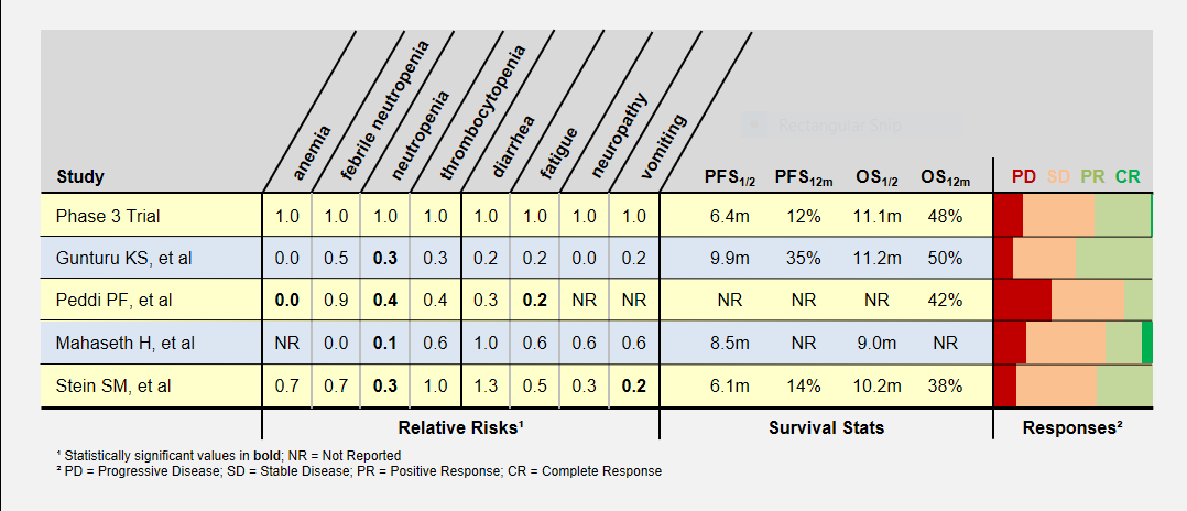
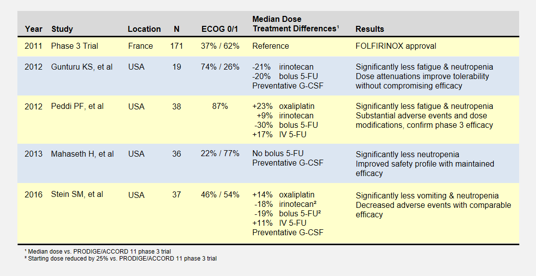
I summarize four studies that report on efficacy and side effects of low dose FOLFIRINOX in metastatic pancreatic cancer. The following table summarizes these studies and the phase III PRODIGE 4/ACCORD 11 approval trial.

Overview of Low-Dose FOLFIRINOX studies.

Overview of Low-Dose FOLFIRINOX Studies

I’ve reported the median dose levels, when available, as compared to the phase 3 FOLFIRINOX clinical trial. Many patients did not start at a full dose, and most had dosage reductions sometime during each study.

About the Studies

The Gunturu KS, et al2 retrospective review included Yale University previously-treated patients from June 2010 to July 2011, documenting their doses, toxicities, and survival results. Reduced doses were not by design, but rather a result of physician discretion. All patients received preventative G-CSF (i.e. neupogen) to prevent neutropenia. This study had a high percentage of healthiest patients.

The Peddi PF, et al3 retrospectively reviewed the FOLFIRINOX experiences of Washington University, the Mayo Clinic, and the University of Wisconsin to compare US “real-world” experiences to the phase 3 clinical trial held in France. Physicians reduced doses at their discretion, not by study design.

Mahaseth H, et al4 retrospectively reported on Emory University’s modified FOLFIRINOX regimen that omitted bolus 5-FU and administered G-CSF to all patients.

Yale University later conducted the Stein SM5, et al prospective study after the promising results in the Gunturu KS study. Starting Irinotecan and bolus 5-FU doses were reduced by 25%, and further at physician discretion. This phase 2 clinical trial (NCT01523457) may provide the most rigorous results.

Patient Responses

I’ve summarized these study’s adverse patient reactions and tumor response statistics in the table below. Relative Risks of greater and less than 1.0 indicate that the study recorded more or less of a particular event, respectively, compared to the phase 3 clinical trial. As an example, a relative risk of 0.3 means that particular event happened 30% as often as in the phase 3 clinical trial. Bold numbers indicate statistically significant findings (p-value < 0.05). Because of the small study sizes, many promising findings did not reach statistical significance.

Patient responses in Low-Dose FOLFIRINOX studies.

Patient Responses in Low-Dose FOLFIRINOX Studies

Progression-Free (PFS) and overall survival (OS) were similar in all studies (the accurate statement is that they were “not found to be different”). Toxicity levels were almost uniformly lower in all low dose FOLFIRINOX studies.

Colored bars represent tumor responses to treatment, with width corresponding to patient counts. Depending upon an individual’s treatment goals, you may want to know different results. For instance, a patient needing to shrink a tumor for surgery may want to look at the PR+CR result. A patient desiring long-term stability may want to minimize the PD result.

Blood-Related Adverse Events

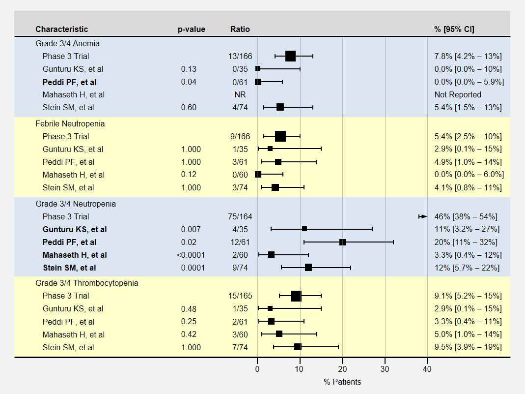
The table below contains more detail and 95% confidence intervals on blood-related adverse events. Studies with fewer participants have less certain results and wider confidence intervals. For adverse event analysis, I included both locally advanced and metastatic patients from each study.

Blood related adverse events of Low-Dose FOLFIRINOX Studies.

Blood Related Adverse Events in Low-Dose FOLFIRINOX Studies

Most adverse events for lower dose FOLFIRINOX were less than the baseline phase 3 clinical trial. However, except for neutropenia, sample sizes or adverse effects were too small to show statistical significance.

Note that the phase 3 clinical trial’s neutropenia rates were especially high. Three studies (Gunturu, Mahaseth, and Stein) used C-GSF for all participants which likely reduced the rate of neutropenia. The Peddi study used C-GSF at higher rates than the phase 3 trial, also reducing the neutropenia rates.

Non-Blood Related Adverse Events

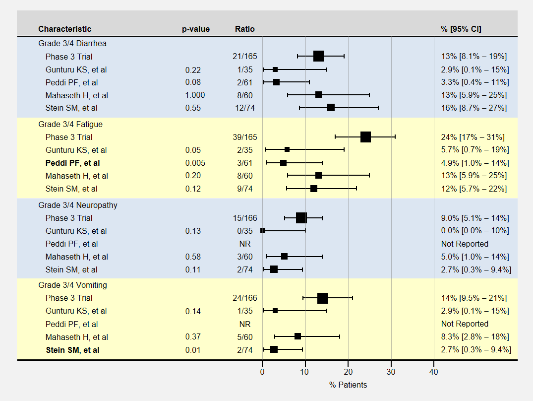
The table below contains more detail and 95% confidence intervals on non-blood-related adverse events. For adverse event analysis, I included both locally advanced and metastatic patients from each study.

Non-blood related adverse events of Low-Dose FOLFIRINOX studies

Non-Blood Related Adverse Events in Low-Dose FOLFIRINOX Studies

Here again, most adverse events for lower dose FOLFIRINOX were less than the baseline phase 3 clinical trial, with sample sizes usually too small to show a statistically significant effect. The Peddi study had significantly less fatigue, and the Stein SM study with a 25% reduction in the initial dose of irinotecan and bolus 5-FU had significantly less vomiting.

How Low Can We Go?

Let’s take a look back at the phase 1 dose escalation trial for FOLFIRINOX6. The study establisged a MTD for oxaliplatin and irinotecan at 85mg/m2 delivered over 120 minutes and 220mg/m2 delivered over 90 minutes respectively. The reduced the recommended level to 85/180 mg/m2 when they realized the cumulative effect would not allow patients to maintain a 2 week schedule.

The dose escalation started with lower oxaliplatin/irinotecan doses as shown in the following table. Important to this discussion, almost every dose level showed anti-tumor activity – including complete responses at the two lowest levels. Participants experienced no dose-limiting toxicities at the three lowest dose levels.

Patient responses in phase 1 FOLFIRINOX dose escalation study

Patient Responses in Phase 1 FOLFIRINOX Dose Escalation Study

The primary goal to determine the dose-limiting toxicity, not effectiveness, opened enrollment to 41 patients with 8 different types of cancers. Of the 6 pancreatic cancer patients, 1 enjoyed a complete and another a partial response (dose levels not specified).

Summary

These peer-reviewed, published studies proved evidence of low dose FOLFIRINOX efficacy similar to the phase 3 trial results. Positive or negative differences in progression-free or overall survival rates cannot be seen with the small studies thus far.

Even with these small study sizes, we have statistically significant evidence of a reduction in toxicity with the low dose FOLFIRINOX regimens.

In the FOLFIRINOX dose escalation trial, patients at the two lowest oxaliplatin/irinotecan levels recorded complete responses with no limiting side effects.

Low dose FOLFIRINOX is better tolerated with significant anti-tumor activity. Oncologists should consider this regimen as an option for patients desiring efficacy but unwilling to endure severe toxic events. Patient experiences with low dose FOLFIRINOX will produce more retrospective studies that will help pinpoint a most effective dose.

I propose that oncologists start patients with low dose FOLFIRINOX and in the absence of severe side effects, increase the dose. The first rounds are often the most difficult as a patient must quickly learn how to deal with chemotherapy-induced nausea and fatigue in addition to their new cancer diagnosis.

References

[1]Conroy T, et al. (2011 May 12) “FOLFIRINOX versus gemcitabine for metastatic pancreatic cancer”. N Engl J Med 364(19):1817-25 PMID: 21561347.

[2]Gunturu KS, et al. (2013 Mar) “FOLFIRINOX for locally advanced and metastatic pancreatic cancer: single institution retrospective review of efficacy and toxicity”. Med Oncol 30(1):361 PMID: 23271209.

[3]Peddi PF, et al. (2012 Sep 10) “Multi-institutional experience with FOLFIRINOX in pancreatic adenocarcinoma”. JOP 13(5):497-501 PMID: 22964956.

[4]Mahaseth H, et al. (2013 Nov) “Modified FOLFIRINOX regimen with improved safety and maintained efficacy in pancreatic adenocarcinoma”. Pancreas 42(8):1311-5 PMID: 24152956.

[5]Stein SM, et al. (2016 Mar 29) “Final analysis of a phase II study of modified FOLFIRINOX in locally advanced and metastatic pancreatic cancer”. Br J Cancer 114(7):737-43 PMID: 27022826.

[6]Ychou, et al. (2003 Mar) “An open phase I study assessing the feasibility of the triple combination: oxaliplatin plus irinotecan plus leucovorin/ 5-fluorouracil every 2 weeks in patients with advanced solid tumors”. Ann Oncol 14(3):481-9 PMID: 12598357.

Long-Term Pancreatic Cancer Survivors Without Surgery

Overview

Curative resection is not offered to most pancreatic cancer patients and information about long-term survivors in this group is scarce. In late 2015, Virginia Mason Medical Center (VMMC) authors documented the characteristics of their eleven long-term pancreatic cancer survivors that never had curative surgery.

Here I’ll present relevant insights into these patients that succeeded at long-term survival. I’ll break down and summarize important information from this paper. Perhaps you will find yourself in one of these patients? Keep in mind that these patients were all treated at VMMC and their therapies will be similar to VMMC’s protocol.

PaperRare long-term suvivors of pancreatic adenocarcinoma without curative resection
AuthorsOh SY, Edwards A, Mandelson MT, Lin B, Dorer R, Helton WS, Kozarek RA, Picozzi VJ
PublicationWorld J of Gastroenterol, 2015 Dec 28; 21(48): 13574-13581
Links[Abstract] [PDF]

Key Points for Patients

  • This paper documents 11 long-term pancreatic adenocarcinoma survivors treated at VMMC (Seattle, WA) who never had curative surgery
  • At diagnosis, all long-term survivors were fully active (ECOG status 0), had normal albumin levels, and tumors at the head of the pancreas
  • 2% [95% CI: 1.0% – 3.5%] of VMMC’s non-resected patients from 1995 to 2009 were ≥5-year survivors
  • All non-metastatic patients were treated with some type of combined chemoradiation
  • Most long-term survivors were overweight which the authors indicate may confer a survival advantage with diseases associated with wasting
  • Long-term survivors were less likely to develop distant metastases with peritoneal metastases being the most survivable
  • Of the 6 deaths recorded, 4 were not directly related to the tumor
  • These patients were treated with VMMC’s preferred treatments. This report does not indicate superiority of their treatment regimen. Data from other institutions would be needed for valid comparisons.

Introduction

Very few publications concerning long-term pancreatic adenocarcinoma (PDAC) survival concentrate on patients who could not have curative resections. A 1988 Gastrointestinal Tumor Study Group paper suggested that long-term survival without surgery might be possible for some patients.1 A 2014 Korean review of 482 inoperable PDAC patients identified characteristics of long-term survivors, defined as >18 months for stage 4 and >27 months for stages 2-3.2 However, the paper I review here provides much more clinical detail on 11 long-term (≥5-year) pancreatic adenocarcinoma survivors treated at VMMC in Seattle, WA who never had curative surgery.

Virginia Mason Medical Center Pancreatic Cancer Patients (1995-2009)

Virginia Mason Medical Center Pancreatic Cancer Patients (1995-2009)

The Long-Term Survivors

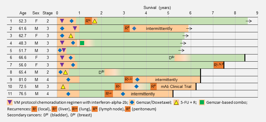
The table below summarizes the VMMC’s long-term pancreatic cancer survivors diagnosis and treatment histories with additional details in the paper. At publication time, five long-term survivors survived. Of the remaining six, four died from sepsis not directly related to their tumor and the two metastatic patients died from disease progression.

The authors suspect these patients happened to have unique tumor biology that was susceptible to these treatments.

Treatment and survival details of VMMC's long-term survivors without curative surgery

Treatment and survival details of VMMC’s long-term survivors without curative surgery

Common Attributes

All long-term pancreatic cancer survivors were in excellent health upon diagnosis. All had tumors in the head of the pancreas. Most were overweight or obese, a risk factor for pancreatic cancer, but which may also be a survival advantage as wasting is a common symptom.

Radiation

VMMC treated all locally advanced long-term pancreatic cancer survivors with some form of radiation. Most received the Virginia Mason protocol consisting of interferon-alpha-2b, 5-FU, cisplatin and radiation while the others received chemoradiation consisting of 5-FU and radiation (one had both). Radiation use is controversial and its importance in long-term pancreatic cancer survival is unknown.

Neither metastatic long-term pancreatic cancer survivors was treated with radiation.

Chemotherapy

As you might expect, Virginia Mason gave all long-term pancreatic cancer survivors their preferred treatments for pancreatic adenocarcinoma – especially the VM protocol and Gemcitabine/Doxetaxel. These treatments are not commonly used elsewhere. We won’t understand the importance until other institutions publish details about their long-term pancreatic cancer survivors using their preferred treatments. I suspect that a different set of their patients would thrive if they used another regimen.

Genetics

Patient #5 had a family history of cancers and a BRCA2 mutation. Due to other considerations, treatment lasted only 4 months but was unusually effective. The authors note that the VM protocol uses cisplatin which has been effective against BRCA2 tumors.3

Tumor Progression

In long-term survivors, tumors commonly progressed only in or near the tumor bed (local, lymph node, or peritoneum).

Conclusion

This paper gives a rare look at long-term pancreatic cancer survivors without curative surgery. Long-term survivors had excellent health, pancreas head tumors, local or no metastases, and effective treatments with long treatment holidays. Stenting of liver ducts was often necessary.

These patients were given VMMC’s preferred treatments. This report does not indicate superiority of their treatment regimen. Data from competitive institutions is needed for valid comparisons. I would not focus on the specific VMMC treatments.

The findings I find important are:

  • first-line treatment was effective
  • second-line treatment was effective
  • distant metastases rarely appeared
  • maintenance of body weight

Long-term survival of pancreatic cancer is possible. I believe that we need Investigation into matching treatments to patients to improve outcomes for more patients.

Read the paper for additional details.

Discrepancies

Here I note discrepancies between the paper and my interpretation.

  • The paper says: “Non-resected ≥5-year survivors represented 2% (11/544) of all non-resected patients undergoing treatment for pancreatic cancer…”. By my count, there were 555 (544 <5-year survivors + 11 ≥5-year survivors) total patients. It does not change the 2% ratio, but I used 555 total patients when calculating the 95% Confidence Interval reported here.

Driving treatment decisions on 28 Aug 2016

References

[1]Gastrointestinal Study Group. (1988). “Treatment of locally unresectable carcinoma of the pancreas: comparison of combined-modality therapy (chemotherapy plus radiotherapy) to chemotherapy alone”. J Natl Cancer Inst 80(1):751-755. PMID: 2898536.

[2]Jo JH, et al. (2014 Oct). “Clinical characteristics of long-term survivors of inoperable pancreatic cancer: an 8-year cohort analysis in Korea”. Pancreas 43(7):1022-31. PMID: 24991970.

[3]Vyas O, Leung K, et al. (2015). “Clinical outcomes in pancreatic adenocarcinoma associated with BRCA-2 mutation”. Anticancer Drugs 26:224-226. PMID: 25304989.

Cancer Stem Cell Model

Driving treatment decisions on 10 Nov 2015

Scientists use cancer models understand cancer. These models describe the rules cancer follows – how it starts, grows, metastasizes, and ultimately how it can be killed. Reviews of models generate new treatment ideas that models indicate should be successful.

Models of how cancer works drive the direction of cancer funding, research, prevention and treatment decisions. Faulty models lead to research producing ineffective treatments. Some researchers say that is happening now.

This post introduces the cancer stem cell model. The cancer stem cell model is not widely accepted but is gaining traction and a share of research money. It was developed as an alternative to the clonal evolution model to explain treatment failures.

Cancer Stem Cell Model

The cancer stem cell (CSC) model is an alternate model to explain the origin and diversity of cancer – and why past treatments have failed2. This model says that some (perhaps all) cancers are driven by a small number of treatment-resistant cancer cells with stem cell-like properties3,5. Stem cells have a slower life cycle and thus are largely unaffected by traditional chemotherapies that disrupt rapidly-dividing cells.6

Cancer Stem Cells Accumulate Mutations

Cancer Stem Cells Accumulate Mutations

[The Cancer Stem Cell model was hard to research. There are many papers and web seminars that present their research as, “this is the way it is – no disputes”. Sorting out what is commonly accepted or not takes a lot of review and I’m certain I don’t have it all correct. This is the best I’ve come up with.]

A cancer stem cell is very long-living, can accumulate genetic mutations over its lifetime, and then produce a nearly unlimited supply of cancer cells containing these mutations. Just as in the clonal evolution model, these cancer cells could continue to generate new mutations and divide uncontrollably.

[Whether CSC’s are actual adult stem cells that have become cancerous or are normal cells that have acquired “stem-like” properties is under active investigation.]

Tumor Growth under the Cancer Stem Cell Model

Tumor Growth under the Cancer Stem Cell Model

The key to the cancer stem cell model though, is the small colony of cancer stem cells (red cell above) that regenerate cancer cells but are killed differently than normal cancer cells.5 Killing off the stem cells will result in the eventual dissipation of the tumor as it can no longer regenerate.

Cancer Stem Cell Model on Chemotherapy

Cancer stem cell model response to chemotherapy treatments

Cancer stem cell model response to chemotherapy treatments

Accumulate Mutations

In the CSC model, a long-lived stem cell accumulates the cancer-causing mutations. It is believed that the property of a long lifetime allows it to accumulate all these mutations. A normal cell, with its much shorter lifespan, would be unlikely to accumulate enough mutations. The key difference is that at the core of the tumor is a CSC, sometimes called a tumor-initiating cell.

[I’m couching the discussion with phrases above like “it is believed”, but if you read the stem cell theory papers, these expressions of doubt are not presented. Some stem cell theorists say that a normal cell gains mutations and becomes stem cell-like to drive the cancer.]

Tumor Growth

The CSC is the major producer of all the new cancer cells3. The normal cancer cells (NCS) have limited cell division capability (just like normal cells). The CSC can continue to mutate which also results in tumor heterogeneity.

Treatment

As treatment begins, susceptible cells are again destroyed. However, CSC’s, which are slowly dividing, are not susceptible to chemotherapy3,5. To make matters worse, the CSC’s, acting like stem cells, see the tumor’s tissue damage and do what all stem cells do – regenerate new tissue (more CSC’s!). The result could again be a smaller tumor, but with an even larger concentration of CSC’s. In this model, what looks like good news on a CT scan (a smaller tumor) is really bad for the future.

[From a patient perspective, the CSC and clonal evolution model behave the same on scans, but the resulting tumor is very different. If the CSC model is correct, then in the long run, giving treatments that don’t kill the CSC’s is a bad thing to do.]

Treatment Holiday

After treatment, driven by many more CSC’s, the tumor growth accelerates.

[Question for Researchers: How does this square up with patients who have long-lasting remissions to chemotherapy?]

Cancer Stem Cell Model on Chemotherapy and Stem Cell Therapy

Under the CSC theory, the correct treatment protocol is to target the CSC’s themselves. Forget about the other cancer cells. Once the CSC’s are gone, the normal cancer cells cannot keep going by themselves and eventually perish. You can use a normal chemotherapy agent in addition to the CSC treatment in order to hasten the demise.

Cancer stem cell model response to chemotherapy and stem cell treatments

Cancer stem cell model response to chemotherapy and stem cell treatments

Treatment

As treatment begins, susceptible cells are again destroyed. In theory, stem cell therapy eliminates the CSC’s. Careful targeting of the CSC’s must be done to make sure that normal stem cells are not affected – which would probably be devastating to the patient.

Treatment Holiday

After treatment, with no CSC’s to replenish them, the normal cancer cells eventually die off.5

Controversy

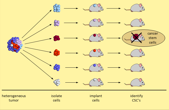
The cancer stem cell model is not embraced by many cancer experts. The primary evidence is based on a set of experiments that break a tumor down and separate the tumor cells into different types. When 20,000 (or so) tumor cells of one type are transplanted into mice, a tumor does not take hold. When just 200 tumor cells of another type are transplanted, the tumor grows. This second set of tumor cells are considered CSC’s because they initiated human cancer growth in the mice. Experiments like these identify tumor cells with stem-like properties.

Identifying Cancer Stem Cells

Identifying Cancer Stem Cells

Skeptical researchers say that a demonstration of human tumor cell growth in immunodeficient mice is insufficient. Growing human cancer cells in a mouse is too dissimilar an environment to provide proof that these are cancer stem cells.5

It should also be noted that testicular cancer that is curable with chemotherapy alone. If cancer stem cells were involved, this would not be possible. So apparently CSC’s do not drive all cancers.

Another View of the Cancer Stem Cell Model

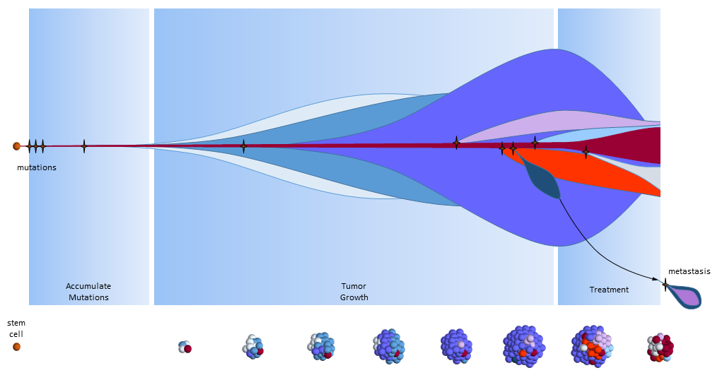
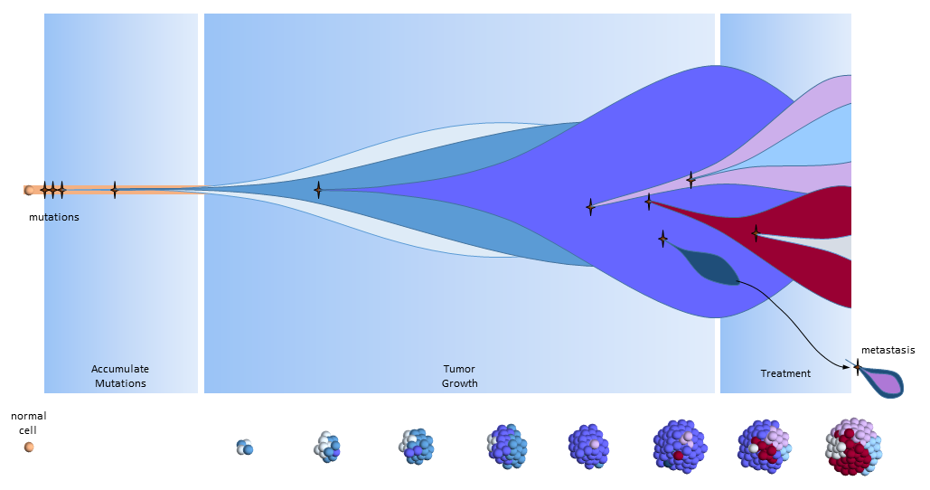
The figure below presents another way to look at tumor growth in the cancer stem call model. Each colored area represents a cell colony with a specific set of mutations. Time progresses to the right. The height of each colored area represents the quantity of cells in the colony. New mutations are represented by stars and may originate from any established colony. The figure shows that these new mutations only originate from the tumor’s cancer stem cells (dark red) and then compete for space and resources with other colonies.

Chemotherapy treatment is effective on the normal cancer cells, but has the opposite effect on cancer stem cells which multiply in response to the tissue damage. After treatment ends, a larger number of cancer stem cells are present to begin tumor regrowth.

Eventually, one of the colonies acquires the ability to metastasize and migrates to another organ.

Cancer Stem Cell Model with Effective Chemotherapy

Cancer Stem Cell Model with Effective Chemotherapy

Summary

The cancer stem cell model is an alternate explanation for tumor growth and response to treatments. It was devised to try to explain failures of treatments based on the correctness of the clonal evolution model. The correctness of the cancer stem cell model is hotly debated.

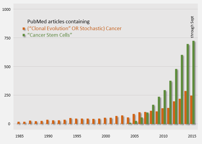
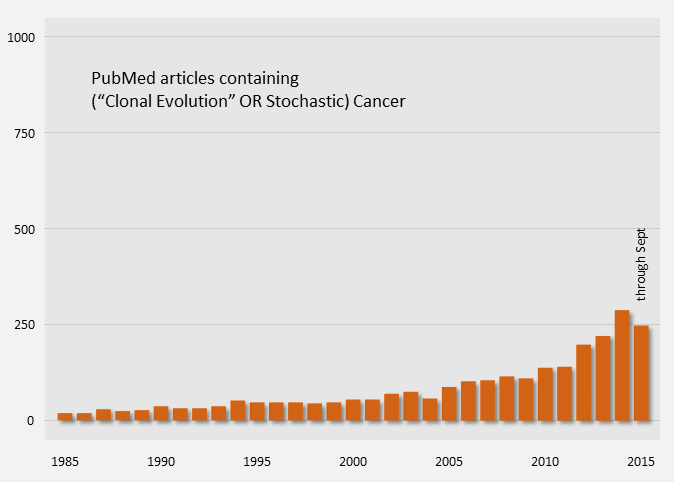
Publication counts of Cancer Stem Cell papers started in 2004 and have been on a steep rise, indicating that it is an active topic. For comparison, publication counts for the clonal evolution model are shown in orange below.

Cancer Model Publication Counts (PubMed)

Cancer Model Publication Counts (PubMed)

Assuming that the cancer stem cell model is true, here are some points to consider.

  • CSC’s are rare, “immortal” cells found in some cancers
  • CSC’s are a tiny fraction of the total tumor (<1%)
  • Radiotherapy and chemotherapy kill off normal cancer cells, but CSC’s respond by multiplying
  • When normal cancer cells are depleted, CSC’s regenerate new cancer cells3,6
  • The only cure is complete elimination of the CSC’s:
    • Surgical resection
    • Stem-cell targeting treatment (experimiental), likely combined with traditional treatments4,6

Surgical resection of solid tumors (before metastases) remains the only curative treatment common to both models.

References

[1] Nowell PC (October 1976). “The clonal evolution of tumor cell populations”. Nature 194(4260):23-8. PMID: 959840.

[2] Soltysova A, Altanerova V, et al. (2005). “Cancer stem cells”. Neoplasma 52(6):435-40. PMID: 16284686.

[3] University of Michigan Cancer Stem Cell Research Introduction web page (accessed 21 Sep 2015) http://www.mcancer.org/research/stem-cells/introduction

[4] University of Michigan Cancer Stem Cell Research Treatment web page (accessed 21 Sep 2015) http://www.mcancer.org/research/stem-cells/introduction/treatment-options

[5] EuroStemCell web page: Cancer: a disease of stem cells? (accessed 21 Sep 2015) http://www.eurostemcell.org/factsheet/cancer-disease-stem-cells

[6] Science in School web page: Cancer stem cells – hope for the future? (accessed 21 Sep 2015) http://www.scienceinschool.org/2011/issue21/cscs

Clonal Evolution Model

Driving treatment decisions on 5 Nov 2015

Scientist use cancer models to understand cancer. These models describe the rules cancer follows – how it starts, grows, metastasizes, and ultimately how it can be killed. Reviews of models generate new treatment ideas that models indicate should be successful.

Models of how cancer works drive the direction of cancer funding, research, prevention and treatment decisions. Poor models lead to research producing ineffective treatments.

This post looks at the dominant cancer model used by researchers today.

[I became interested in cancer models because each model has its own ideas about the most effective treatments. Treatments suggested by this model are sometimes the absolute wrong treatment under another model. So I began to look into the differences in their recommendations. I wanted to find overlap – treatments that would work well under any model and thereby improving my odds of successful treatment. The importance of surgical resection as a cure stands out very clearly.]

Clonal Evolution Model

You are probably familiar with the dominant cancer model, the clonal evolution model, first proposed in 19761 where a single cell eventually gains enough mutations to become a full-fledged cancer tumor cell. This cancer cell continues to divide uncontrollably to form a tumor mass.

Clonal Evolution Model

Mutation Accumulation Under the Clonal Evolution Model

As the tumor grows, defects in the replication process cause “daughter” cells to add more mutations. Following evolutionary rules, cells that compete better for blood and nutrients become more plentiful. The result is a tumor mass that contains cells with various colonies of mutations dominated by colonies that proliferate best.

The resulting tumor is considered by researchers to be a heterogeneous collection of tumor cells2. This means that all tumor cells are not identical – they contain unique sets of mutations. It is thought that some of the earliest mutations will be present throughout the tumor. Mutations occurring in sections of DNA not used by these cells are called passenger mutations and don’t appear to be harmful3. But mutations that directly result in cells becoming cancerous are called driver mutations. Finding and targeting a driver mutation should lead to effective treatments. Ideally we would like to target a driver mutation that is present throughout the entire tumor.

“Precision medicine” treatments rely on genetic testing from biopsies of tumors. Unfortunately, these biopsies only grab a portion of the tumor and this sample may not be representative of the entire tumor. If a targeted therapy is identified, it may only be effective on the sampled section of the tumor. Tumor cells without the target may be unaffected and continue growing. Effectiveness depends upon “luck of the draw” in finding the right driver mutation.

[It is here where I think the importance of germline genetic testing comes in. This is look for inherited cancer-causing mutations from a sample of saliva or blood. If one of these mutations is found, I think it has a good likelihood of being a driver mutation present throughout the entire tumor. My BRCA2 mutation was found this way and directly led to my platinum-based chemotherapy treatment that reduced the tumor size by 80% (diameter) and enabled surgery.]

Tumor Growth under the Clonal Evolution Model

Tumor Growth under the Clonal Evolution Model

As the tumor continues to grow, eventually mutations will develop that allow tumor cells to metastasize and spread to distant organs.

For over 30 years we’ve been developing new treatments based on this model. Treatments are reducing the size of tumors, but complete elimination without relapse is uncommon. The lack of success has some researchers looking at different cancer models.

Clonal Evolution Model on Chemotherapy

Experience has shown that chemotherapy is usually only effective on a portion of the tumor. Its application can shrink the tumor and change the types of mutations prevalent.

The figure below describes how the tumor grows and responds to chemotherapy. When the tumor size is small (below the dashed line) it may cause adverse side effects, but it is undetectable by scans. This phase can last years and patients typically don’t know they have cancer while the tumor slowly grows in size.

Clonal evolution model response to chemotherapy treatments

Clonal evolution model response to chemotherapy treatments

Accumulate Mutations

In the clonal evolution model a normal cell accumulates mutations as it divides. After enough mutations have accumulated, the cell becomes cancerous. My post The Hallmarks of Cancer describe the cell characteristics these mutations provide for the formation of cancer.

Tumor Growth

The original tumor cell divides uncontrollably and its “offspring” create additional mutations, as indicated by the different colored cells. Some offspring grow and divide better than others. This is evolution on a very rapid time frame. In cancers cells, mutations happen much more quickly than in normal cells because of the loss of DNA repair mechanisms. Most mutations don’t survive, but some do well – even better than previous tumor cells. This is survival of the fittest within the tumor.

The tumor is said to be heterogeneous, meaning it is composed of cancer cells with many different kinds of mutations. In the figure above, tumor cells with different sets of mutations are shown as cells with different color. Tumor heterogeneity has important negative implications for genetically targeted therapies (i.e. precision medicine).

Treatment

After the tumor is large enough to be detected, treatment begins. Cell mutations continue to happen and new treatment-resistant cells begin to proliferate while the others are destroyed. In this phase, the tumor may grow or shrink depending upon how well the tumor cells resist and adapt to the treatment.

In the figure above, the blue and pink colonies are susceptible and killed off by the chemotherapy. A new colony of mutated cells (light blue) are unaffected by treatment and grow rapidly. But overall, the tumor shrinks below the size of detection where the patient is then considered to be in remission and treatment is halted. Patients may assume that the tumor is completely gone, but most likely it is present but too small to detect (remission, not cured).

Colonies of cells susceptible to treatment make up smaller and smaller portions of the tumor. Treatment effectiveness comes to a halt when the susceptible cells have been killed, leaving behind the treatment-resistant colonies.

[Treatment targeting a driver mutation may be much more effective – even resulting in a long-term remission. Unfortunately, because of the tumor’s ability to mutate to readily, a mutation may come that works around even this driver mutation. Evolution is a very powerful process and is working in the tumor’s favor. Surgical resection before it gets the chance to mutate again may still be the surest cure.]

Treatment Holiday

After treatment is halted, tumor cell growth of remaining cells continues unrestrained. By this time, the tumor may be composed of completely different types of tumor cells, ones that are largely unaffected by previous treatments.

What remains is a treatment-resistant tumor. Another treatment must be selected. A new biopsy might identify another treatment that will also likely treat a portion of the tumor.

Another View of the Clonal Evolution Model

The figure below presents another way to look at tumor growth in the clonal evolution model4. Each colored area represents a cell colony with a specific set of mutations. Time progresses to the right. The height of each colored area represents the quantity of cells in the colony. New mutations are represented by stars and may originate from any established colony. The figure shows that these new mutations can originate in any part of the tumor and then competes for space and resources with the others.

Eventually, one of the colonies acquires the ability to metastasize and migrates to another organ.

Clonal Evolution Model

Clonal Evolution Model

Summary

The clonal evolution model of cancer is the widely accepted explanation for tumor growth and response to treatments. Publications on the topic have been ongoing for quite some time.

Clonal Evolution Model Publication Count

Clonal Evolution Model Publication Count

The clonal evolution model describes how cancer develops from a single cell to become a tumor, becomes heterogeneous, responds to treatment, and becomes resistant to treatments.

The model points to a few effective treatment options for patients.

  • Surgical resection before metastases
  • Treatments targeting driver mutations

Much current research and development is being done to target mutations found in tumors. The next model I’ll discuss says that we’re targeting the wrong mutations and wrong tumor cells.

References

[1] Nowell PC (October 1976). “The clonal evolution of tumor cell populations”. Nature 194(4260):23-8. PMID: 959840.

[2] Heppner GH. (Jun 1984). “Tumor heterogeneity”. Cancer Res. 44(6):2259-65. PMID: 6372991.

[3] Greenman, Chris, et al. (Aug 2006). “Statistical Analysis of Pathogenicity of Somatic Mutations in Cancer”. Genetics 173(4):2187-2198. PMID: 1569711.

[4] Yates LR, Campbell PJ. (Nov 2012). “Evolution of the cancer genome”. Nat Rev Genet. 13(11):795-806. PMID: 3666082.

Cancer Models

Driving treatment decisions on 25 Sep 2015

Cancer models are used by scientists to define and better understand cancer. They describe the rules cancer follows – how it starts, grows, metastasizes, and ultimately how it can be killed. Reviews of their cancer models generate new treatment proposals that the models indicate should be successful.

Cancer models are driving the direction of cancer funding, research, prevention and treatment. Faulty cancer models lead to research producing ineffective treatments. Some researchers say that is happening now.

Cancer Models Example

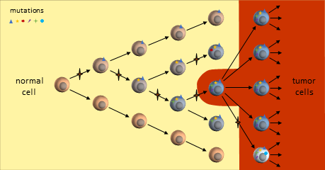
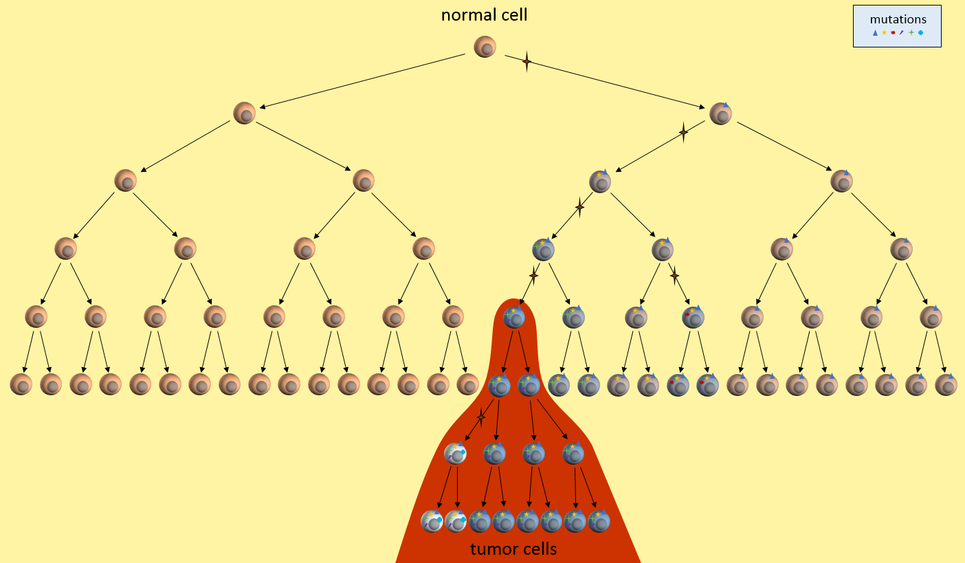
Let’s look at one example of a simple cancer model. It’s just a picture that describes how a normal cell might progress to a cancer cell and then a tumor. In this model, each time the cell divides, a mistake in the replication process results in a mutation (indicated by the star symbol) in one of the two daughter cells. After enough divisions with accumulated mutations, a cancer tumor cell results (highest cell in the red area). All subsequent daughter cells are cancer cells and additional mutations keep happening. The tumor ends up containing cells with varying kinds of mutations. However, the mutations present in the first tumor cell remain present in all daughter cells. These are called driver mutations1, because they drove the initial cancer and will be present throughout the entire tumor.

Model of Mutation Accumulation in the Clonal Evolution Model

Let’s just take this simple model, and ask ourselves some questions. What are this model’s weaknesses? What treatment decisions might this model recommend?

Testing the Model

First, let’s look at what our model implies about cancer growth. Does it fit with observations from real-world patients?

Mutations Only During Replication?

What does our model tell us about cancer growth? For instance, is it reasonable that mutations only happen at during replication? Well, we seem to know that cells need about 20-30 mutations before they can become cancerous. We also know that normal cells can only replicate about 40 to 60 times before their telomere length is too short to allow any more replications. Given this, and DNA’s ability to repair itself, we might have to adjust our model to allow more than one mutation per replication, or allow mutations to happen without replication, or grow longer telomeres. We might design experiments to determine which of these is more correct and then update the model to reflect the new findings.

Are There Nearby Pre-Cancerous Cells?

Our model also seems to indicate that there will be many cells nearby with some mutations, but not enough to yet be cancerous, or pre-cancerous. Is this something that a pathologist can determine? Can we separate out nearby normal-looking cells and genetically sequence them to see if they have some mutations but not all?

Timeline Of Cancer Development?

We might observe that our model seems to require quite a few generations of replications before the first cancerous cell develops. Where to all these cells go? What does this tell us about how quickly cancer could develop? What ages cancers might develop? Does this fit in with the childhood leukemia? Does it fit in with pancreatic cancer? Do we need different models for these cancers? Which cancers does it fit?

Cancer models raise many questions like these that drive directions of research that demand funding. Answering these questions allow us to tweak the model to be more accurate. The more accurate the model, the better its predictive power for new treatments.

Cancer Model Development Flowchart

Cancer Model Development Flowchart

Refining the Model

After we’ve tested the model and experimentally determined new facts, we can update the cancer model. The updated model would be tested again, continually refining the model.

Using the Model

Once we’re happy that the cancer model is mostly reflecting reality and is able to answer basic questions about cancer’s behavior, we are ready to put this model into practical use.

Treatment Decisions

What does our model tell us about treatment decisions? For one, this model seems to indicate that there are certain mutations that will be present in all daughter tumor cells. Examine those nearby pre-cancerous cells. Do they hold the key to uncovering these cancer driver mutations1? If we can find these driver mutations and target them with treatments, we might be able to kill off the entire tumor. This is the idea behind the NIH’s Precision Medicine Initiative.

Tumor Heterogeneity

Our model also says that many mutations will only be present in a part of the tumor – not the entire tumor. See those white and blue cancer cells in the model? They represent tumor cells with different sets of mutations. If we treat only for the white cells’ mutations, the blue cells might continue to grow. This observation fits current patient outcomes.

Early Detection

What does our model say about early detection? Our model indicates that precancerous cells will be around for a while before the cancerous cells develop. It there some way we might be able to detect these pre-cancerous cells? Perhaps they affect the environment around them or put out markers that might be picked up in the bloodstream? This model might drive the development of another model that looks at these issues in the cellular environment.

(Re)-Refining the Model

The cancer model is always being updated to reflect the best knowledge of how cancer works. This simple model really just deals with cancer at a very high level, but even so, it reveals directions for research, early detection, and treatments. Continual refinement of even this simple model brings forth new ideas to be tested, further feeding the models.

Summary

Don’t think that there is some “master” cancer model somewhere that all researchers work from. Each group has its own models developed from their own experience, lab tests, ideas from other groups, etc. These cancer models describe some aspect of cancer as it is understood by the local research team.

In the next posts, I’ll describe two current cancer models. The clonal evolution model2 that has been driving research and funding decisions for several decades. And the cancer stem cell model3 that is not widely accepted but is gaining traction. Which cancer model is (more) correct has a profound effect on patient treatment choices.

Our lack of progress could mean we need to take another look at our basic assumptions, the cancer model, and develop a new model that describes not only how cancer starts, grows, and metastasizes, but also accounts for why we’ve been failing in the “war on cancer”. Each model will undergo continuous revision and tweaking based on new tests to improve their accuracy.

References

[1] Stratton MR (9 April 2009). “The cancer genome”. Nature 485(7239):719-24. PMID: 19360079.

[2] Nowell PC (October 1976). “The clonal evolution of tumor cell populations”. Nature 194(4260):23-8. PMID: 959840.

[3] Soltysova A, Altanerova V, et al. (2005). “Cancer stem cells”. Neoplasma 52(6):435-40. PMID: 16284686.

What Researchers Know and You Don’t

An explanation for what’s driving drug development

In one of the most highly cited cancer papers, The Hallmarks of Cancer1, researchers Doug Hanahan and Bob Weinberg described six common traits of cancer. Their 2011 update, Hallmarks of Cancer: The Next Generation2, added two more traits for a total of eight “hallmarks” of cancer. They also describe an additional two “enabling characteristics” that are not necessary, but if present, hasten the cancer process.

In these papers, they hypothesize that eight specific functions of normal cells must be impaired for them to become cancer cells. A cell could develop these impairments over time, in any order, but only after developing all or most of them could it become a cancer cell.

The hallmarks listed in these papers are understood by every cancer researcher and referenced by thousands of research papers. This is what researchers know and you don’t – yet.

What Does This Mean For Patients?

  • The time needed to accumulate all these impairments explains why cancers are more likely as we age.
  • The requirement to have all these impairments together explains why cancers develop so rarely.
  • Inheriting a mutation of an enabling characteristic explains why hereditary cancers develop in younger people. In essence the deck is stacked against them.
  • A treatment targeting any one of these hallmarks would prevent a tumor from developing.

Implications for Cancer Treatment

The last item above explains a lot about the current direction of cancer drug development. If you were to repair any of these functions, you could potentially stop cancer. Targeted treatments being developed today attempt to repair one or more of these traits in an effort to halt cancer growth.

For example, one hallmark is that cancer cells evade detection by the immune system. This means that cancer cells have developed some way to keep the immune system from recognizing the cancer cells as undesirable. Current immunotherapy drugs are trying to “fix” the immune response to recognize the cancer cells. In pancreatic cancer, treatments currently in later-stage clinical trials that target this hallmark include GVAX vaccine (Johns Hopkins & Aduro BioTech), Algenpantucel-L (NewLink Genetics), CAR T-cell therapy (U of Penn)4.

Patients should note that when cancer cells are targeted in a specific hallmark, they will often develop an alternate method of re-impairing the hallmark function and continue growing as a tumor. For example, there are many ways of deactivating the immune system. Fixing one part of the immune response usually leads to the cancer cells developing an alternate method to halt the immune response. Cancer cells’ ability to continue mutating as they divide means they can eventually stumble on an alternate mechanism. I imagine the tumor as a massively parallel computer that can try thousands of mutation experiments simultaneously where only needs one to succeed at foiling the treatment. Researchers can only target a specific one of these mechanisms at a time.

In the future, perhaps we will combine these therapies in a multi-pronged attack on cancer cells such as is proposed for NSCLC5? Or alternating between two effective treatments to keep cancer from developing resistances? I suspect that this process is at the beginning of a long road.

Hallmarks of Cancer

Here are the hallmarks of cancer in plainer English. The explanation in FutureLearn‘s MOOC Cancer and the Genomic Revolution6 was well presented.

Hallmark1, 2Example Therapeutic Targets2
Growth signals stuck ONEGFR Inhibitors
Ignore anti-growth signalsCyclin-dependent Kinase Inhibitors
No "kill" switchProapoptotic BH3 Mimetics
Unlimited replicationTelomerase Inhibitors
Compels new blood suppliesVEGF signaling Inhibitors
Migrate to other organsHGF/c-Met Inhibitors
Energy production using little O2Aerobic Glycolysis Inhibitors
Deactivate immune systemImmune Activating anti-CTLA4 mAb
Enabling Characteristic2
Easier MutationPARP Inhibitors
Favorable inflammation environmentAnti-inflammatory drugs

I’ll try describing the hallmarks of cancer with an automobile factory analogy. In this analogy, cancer cells are automobiles and uncontrolled growth is accomplished by means of the factory building these autos. These automobiles are often defective, unsafe, but they’re practically free and plentiful. But because of the random defects in these autos, many of them don’t even work at all. But some do and they can have scary defects.

In a more correct analogy, each tumor cell is its own factory producing more defective cars, but we’ll stick with the one factory for now.

The heading for each Hallmark is followed by the name from the original paper in italics and in parentheses. I think you’ll understand why I’ve interpreted them.

Always ON Growth Signals (Sustaining Proliferative Signaling)

One attribute of cancer cells is that they are always replicating, growing the tumor larger and larger.

In our automobile factory, the assembly line never stops. It is working three shifts every day of the year, putting our defective cars out on the road.

Ignore Anti-Growth Signals (Evading Growth Suppressors)

Cancer cells ignore signals that neighboring cells send saying that there’s enough of them and they should stop dividing.

Our dealerships can’t even store all the cars we’re making. They are saying, “Stop! We don’t want any more of your cars!” But their pleas fall on deaf ears. This factory keeps producing cars.

No Kill Switch (Resisting Cell Death)

Cancer cells are made with lots of mistakes in their DNA. Normally, if those defects cannot be fixed, the cell is instructed to kill itself.

Our workers are pretty sloppy and we’re always producing cars with random mistakes. But in our factory, we have no quality control so that all cars are shipped. A lot of them don’t work at all. But some of them do and have some pretty scary attributes. In some, the brakes don’t work. In others, the accelerators are stuck on. In still others, airbags won’t deploy. All defects that should be caught and cause these autos to never see the light of day. But we’ve got no quality control so every car ships.

Unlimited Replication (Enabling Replicative Immortality)

Cancer cells can replicate themselves almost without limit. Normal cells can only replicate up to about 20 times. Their telomeres are shortened by every replication, but cancer cells have found a way to replicate without shortening their telomeres.

In our factory, the robots, machines and tools never wear out or break down. We can produce cars almost without limit.

Compel New Blood Supplies (Inducing Angiogenesis)

Cancer tumors grow very rapidly but still need to be supplied with blood and nutrients. These cells send out signals tricking arteries into branching into the growing tumor.

Our factory managers have bribed their suppliers to provide all the raw materials we need to continue making our cars. They’re not paying them properly and we’ve resorted to trickery to obtain our supplies.

Migrate to Other Organs (Activating Invasion & Metastasis)

Eventually cancer cells develop a way to metastasize. As an example, a cluster of perhaps 100 cells detaches from the tumor, entering the blood stream and gets caught by the liver as it tries to filter out impurities. There it lodges and starts to grow inside the new organ.

This automobile factory becomes so successful at producing cars that the managers decide to open a new factory in another state or country.

Energy Production Using Little O2 (Deregulating Cellular Energetics)

Cancer cells use sugar in a different way than normal cells. For not well-understood reasons, cancer cells have developed a less efficient process to make energy that also uses less oxygen.

Our factory needs lots and lots of energy to make these cars, 24×7. We have access to the same power source that any factory would have, but we also have solar and wind power to supply our energy demands. We’ve developed alternate sources of energy.

Deactivate the Immune System (Avoiding Immune Destruction)

Cancer cells are really normal cells that have mutated to have all the above attributes. The immune system does not recognize them as foreign invaders because they really aren’t – they came from inside our own bodies. Even if the immune system did start to attack, cancer cells have figured out how to distract the immune cells.

Our factory has figured out how to avoid government regulations. We keep producing defective cars, cheating our suppliers, have unapproved power sources, and clearly run afoul of labor laws, but no one comes in to shut us down. Perhaps the factor owners have paid “protection money” to keep regulators looking the other way?

Summary

Articles like The Hallmarks of Cancer provide an insight into the thinking of researchers. Understanding the Hallmarks can help direct you to promising treatments and understand why we’re not at a curative treatment stage yet. Highly mutative tumor cells can bypass any single drug we develop. Eventually we may develop enough targeted treatments that used together will halt cancer growth.

To me, it reinforces the idea that surgical removal is still the best and only pancreatic curative option. Working towards that option will be my primary goal.


References

[1] Hanahan D, Weinberg RA (January 2000). “The Hallmarks of Cancer”. Cell 100 (1): 57–70. doi:10.1016/S0092-8674(00)81683-9. PMID 10647931

[2] Hanahan, D.; Weinberg, R. A. (2011). “Hallmarks of Cancer: The Next Generation”. Cell 144 (5): 646–674. doi:10.1016/j.cell.2011.02.013. PMID 21376230

[3] Scowcroft, Henry (2010). Science blog: http://scienceblog.cancerresearchuk.org/2010/11/10/ncri-conference-the-hallmarks-of-cancer/

[4] Cancer Research Institute’s Pancreatic Cancer web page (accessed 7 Aug 2015) http://www.cancerresearch.org/cancer-immunotherapy/impacting-all-cancers/pancreatic-cancer

[5] Turke, Alexa B, et al. (January 2010). “Preexistence and Clonal Selection of MET Amplification in EGFR Mutant NSCLC”. Cancer Cell 17 (1): 77-88. doi:10.1016/j.ccr.2009.11.022. PMID 20129249

[6] FutureLearn MOOC, Cancer and the Genomic Revolution, University of Glasgow. https://www.futurelearn.com/courses/cancer-and-the-genomic-revolution/